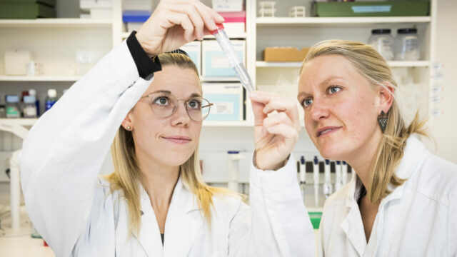

Artificial intelligence is transforming histological assessment by enabling more consistent and quantitative analysis of tissue. At Gubra, our fully digitalized pathology workflow combines whole-slide imaging with AI-powered image analysis to support a wide range of preclinical disease models. We’ve previously explored its application in this blog article series in:
In this article, we focus on ovarian histology and show how AI can automate the assessment of follicles and other key ovarian structures to generate robust, reproducible data.
Why is ovary histology important in preclinical research?
As the field of ovarian biology continues to evolve, the techniques used to evaluate tissue samples for histology must also advance. The reclassification of PCOS (polycystic ovary syndrome) to PMOS (polyendocrine metabolic ovarian syndrome) reflects this development within the women’s health field, emphasizing that the condition extends beyond ovarian morphology alone.
PMOS provides a more holistic and accurate framework by integrating not only alterations in the gonadal–brain axis, but also the substantial metabolic dysregulation, such as insulin resistance and altered energy homeostasis, which is now considered central in the disease pathology.
In parallel, this terminology marks a paradigm shift away from focusing on ovarian “cysts” toward a more precise understanding of the arrested follicles observed in a subset of people with PMOS, thereby bringing structural observations in line with the underlying biology. Evaluation of ovarian tissue is performed in all species during drug development for women’s health diseases, from mouse and rat models to monkey and human samples.
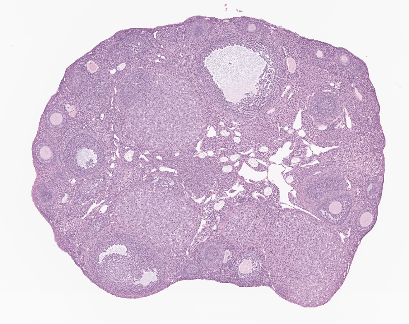

Left: Murine ovary stained with HE. Right: Porcine ovary stained with HE. Both samples demonstrates the diversity of follicle stages and sizes found in the ovary.
In addition to diseases and syndromes that affect the reproductive system, the reproductive system must be assessed during development of pharmaceutical drugs. Preclinical drug development must meet criteria of drug safety and efficacy in rodent models. One such safety end point is to determine if the drug candidate has effects on reproductive tissues, such as the ovary; this sector of preclinical animal work is known as toxicology. To best evaluate the toxicological effects of a drug candidate, histology is performed to evaluate the tissue for signs of oocyte death, follicle defects, or other morphological changes.
Key histological features of the ovary in preclinical models
In preclinical animal models, ovarian histology provides a quantitative window into reproductive health and drug efficacy. A core readout is follicle classification, where follicles are categorized (primordial, primary, secondary, antral, pre-ovulatory, and atretic) based on granulosa cell number and morphology, antrum formation, and oocyte integrity; shifts in this distribution can indicate altered folliculogenesis or accelerated depletion of the follicle pool.
Beyond follicle dynamics, evaluation of stromal fibrosis and inflammation has gained more attention in recent years because of the increased focus on reproductive longevity and ovarian aging. For example, when evaluating fertility preservation treatments, it is essential to assess ovarian stiffness caused by increased collagen deposition, which can result in stromal fibrosis, architectural distortion, and reduced reproductive function. Immune cell infiltration, like an increase in lymphocytes or macrophages within the stroma or around atretic follicles, reflects inflammatory responses that accompany drug-induced or disease-related pathology.
Routine Hematoxylin and Eosin (H&E) staining remains foundational, clearly delineating follicular architecture, granulosa and theca cell layers, corpora lutea, and overall tissue organization. In disease models of primary ovary insufficiency (POI) and PMOS, H&E staining highlights follicular atresia, stromal expansion, or changes to the follicular structures. Complementary histochemical stains such as Picrosirius Red and Masson’s trichrome can be used to visualize collagen and quantify fibrosis. In addition, targeted immunohistochemistry and in situ hybridization technologies like RNAscope™ can further refine interpretation of drug efficacy and increase our understanding of basic and complex biological questions. Collectively, these histological techniques enable both qualitative assessment and quantitative scoring in translational research settings.
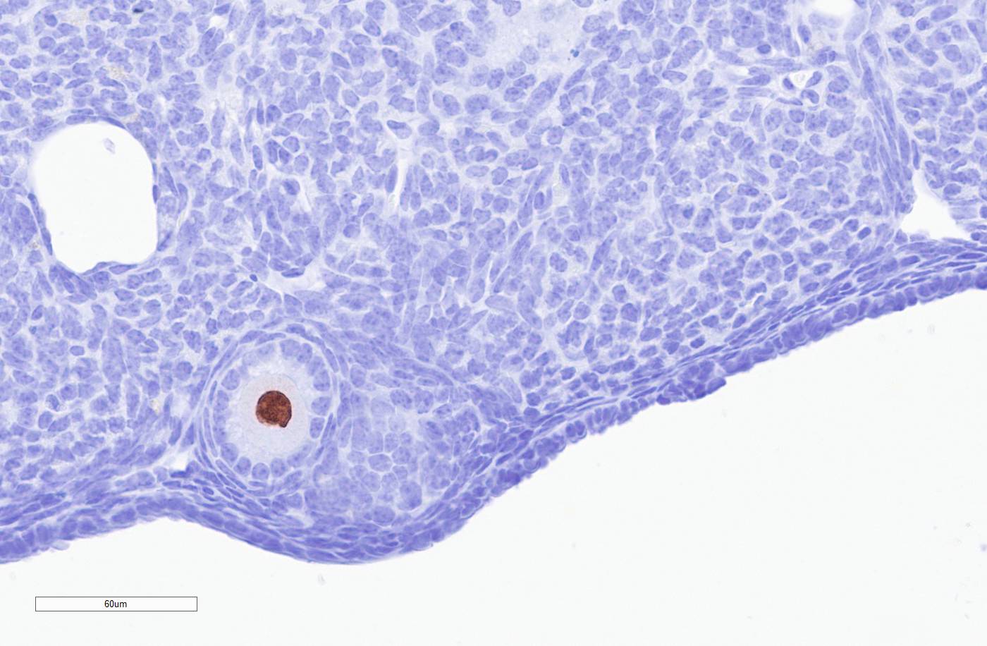
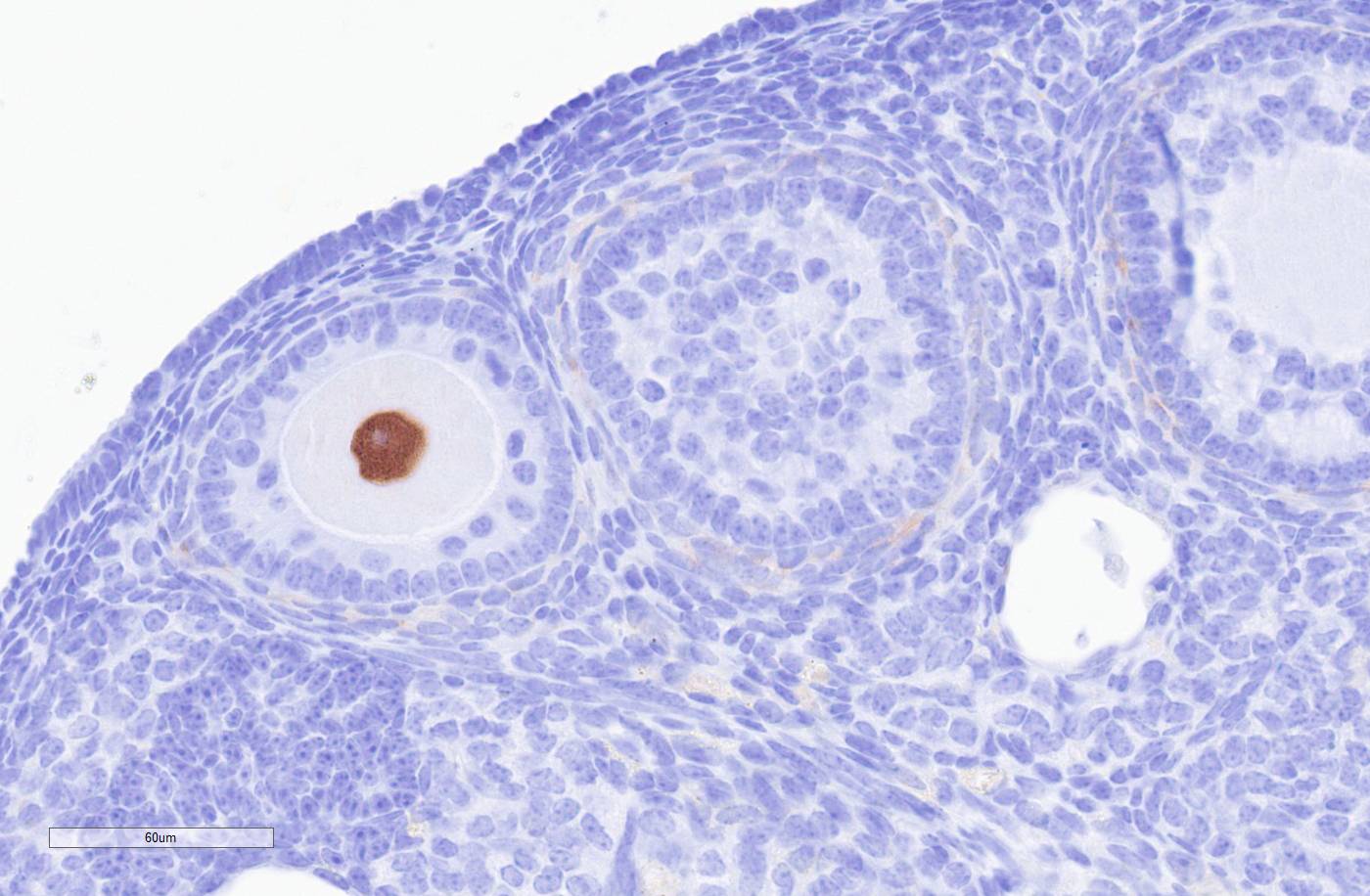
Murine ovary immunohistochemically stained using antibody specific for p63 with follicles with a clearly p63 positive stained oocyte (brown stain).
Quantitative challenges in traditional ovary histology and reproductive endpoints
Traditional ovary histology presents several quantitative challenges that can limit precise assessment of follicle populations and tissue organization. Because analyses rely on thin, two-dimensional sections of a highly heterogeneous three-dimensional organ, follicle counts are susceptible to sampling bias, double counting, or missing structures. Variability in sectioning, staining quality, and subjective follicle classification further contributes to inter- and intra-observer differences, while the labor-intensive nature of manual follicle quantification significantly reduces throughput. Another important, and highly labor-intensive, histological readout is continuous evaluation of estrous cyclicity based on cytological analysis. This method is notoriously low throughput and subjected to inter- and intra-observer differences with manual estrous staging.
Despite these limitations, histological readouts remain critically important, as they provide direct, spatially resolved insight into ovarian architecture, follicle development, tissue health, and overall reproductive function. Thus, while careful interpretation and methodological awareness are essential, traditional histology continues to serve as a cornerstone for understanding ovarian biology.
AI-based quantification of mouse ovary histology
How we use automation and digitalization to increase throughput, robustness and reproducibility of histological endpoints
At Gubra, we regularly perform estrous cycling to determine the estrous cycle stage of our women’s health models. We have fully automated the scoring of the daily vaginal cytology samples using rapid digital scanning and AI models to identify the cell composition based on a H&E staining. This AI model has made estrous cycling not only more efficient, but significantly more reproducible.
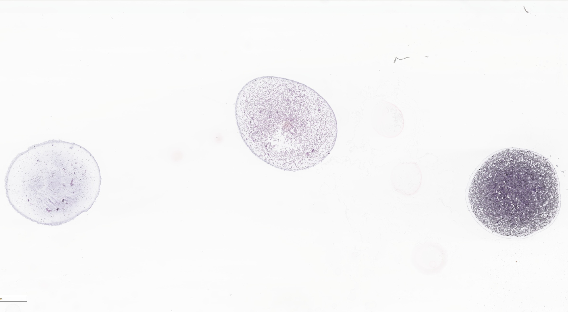
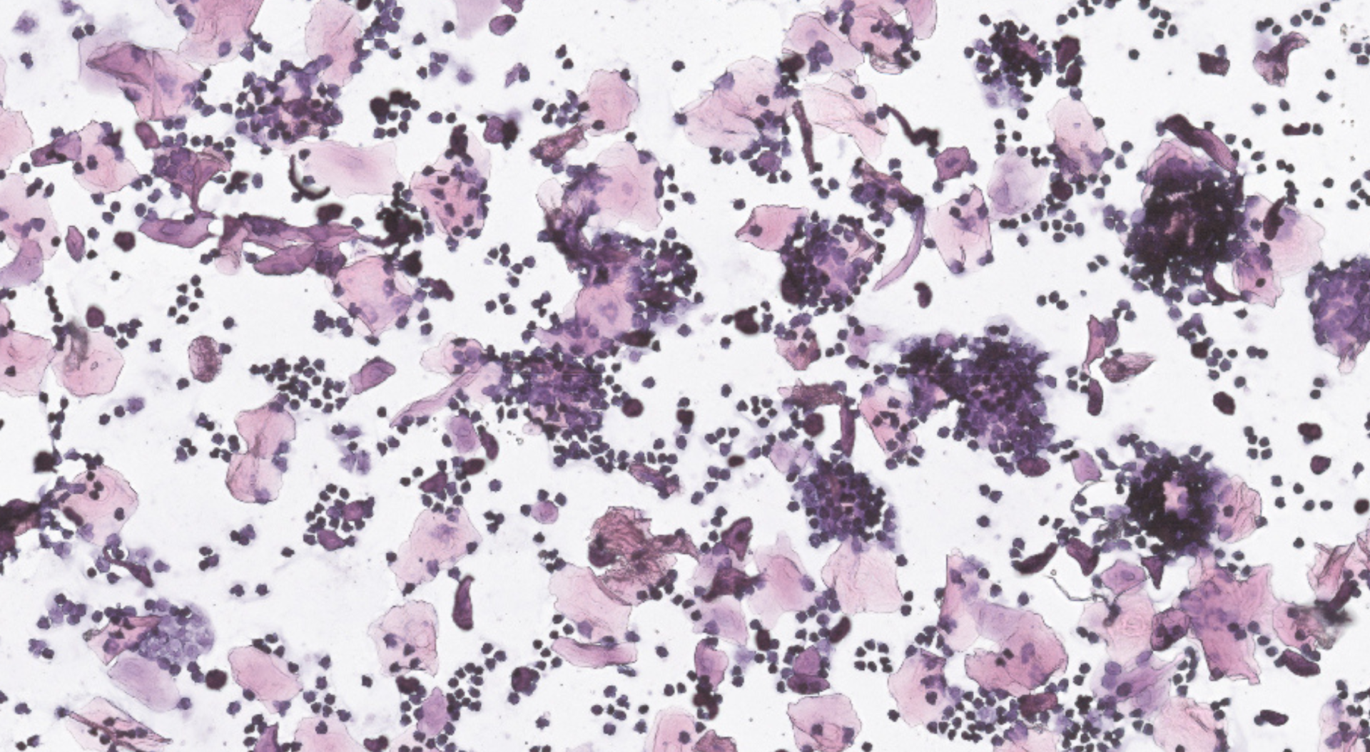
Left: Glass slides with three samples from samples from vaginal lavage. Right: Zoom in of cells in the vaginal lavage samples. Inflammatory and epithelial cells are present.
In addition to our AI-scored estrous cycle samples, we have automated staging of preantral follicles in ovary samples using immunohistochemical staining against p63. We utilize an AI-based algorithm to identify each follicle. Then, upon follicle isolation, the algorithm predicts follicle stage. This algorithm is highly reproducible and performs well when compared to manual scoring by histopathologists. To learn more about this algorithm, see our poster on Automated AI-assisted preantral ovarian follicle staging in a mouse model of chemotherapy-induced primary ovarian insufficiency.
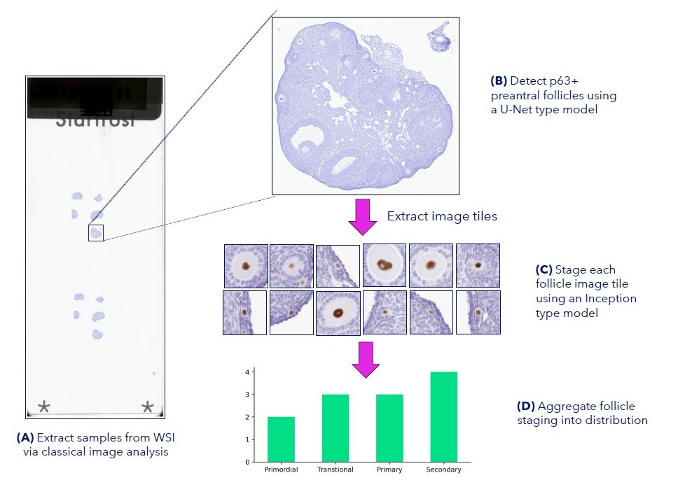
Development of AI-assisted preantral follicle staging analysis pipeline
AI-assisted ovary histology for preclinical women’s health research – Get in touch
AI-assisted analysis is transforming preclinical ovary histology by delivering more consistent, scalable, and quantitative assessment of reproductive tissue and estrous cycle endpoints in animal models. At Gubra, we combine digital pathology, AI-powered image analysis, and deep expertise in preclinical women’s health research to generate robust data that support drug discovery and development. If you’re looking for a CRO with expertise in preclinical women’s health, reproductive disease models, or AI-assisted histology, get in touch to discuss how we can support your next study here.