
Preclinical kidney pathology is transforming as researchers seek more accurate, reproducible, and scalable methods to evaluate disease progression and therapeutic efficacy. Traditional methods are often limited by inter-observer variability, low throughput, and semi-quantitative assessment.
Advances in artificial intelligence (AI) and quantitative image analysis are enabling a new era of digital pathology and AI in histopathology. These technologies allow automated, high-throughput evaluation of histological features such as fibrosis, inflammation, and structural damage, providing deeper, quantitative, and more consistent insights into kidney disease biology.
In this article, we explore how AI-based scoring and quantitative image analysis are advancing preclinical kidney pathology, with a focus on animal models used in chronic kidney disease (CKD), diabetic kidney disease (DKD), and acute kidney injury (AKI).
High-throughput automated and AI-based histological evaluation in preclinical kidney models
The kidney is a complex organ with different structural components, including glomeruli, different tubular segments, and supportive tissue – all organized and located in different zones.
At Gubra we run a fully digitalized image analysis pipeline for quantitative evaluation of tissue samples. This automated image analysis approach enables scalable and reproducible quantitative histology in preclinical kidney models. By applying advanced image analysis techniques, we enable accurate quantification of the cells and lesions identified through histological staining, immunohistochemistry (IHC), or immunofluorescent staining.
In addition to quantification, our platform provides the spatial location of these findings. In the image below, the kidney has been segmented into distinct anatomical zones using AI-based segmentation, making it possible to quantify the marker of interest in the different areas of the kidney.
AI-based kidney compartment analysis
Our advanced AI-powered image analysis enables precise quantification of IHC markers across distinct kidney compartments, including the cortex, outer medulla, inner medulla, and papilla.
By analyzing marker expression within each specific region, we provide a more detailed and compartment-resolved understanding of disease progression and the therapeutic effects of drug treatments. This approach delivers deeper insights into spatial disease patterns, supporting more informed decision-making in preclinical research.
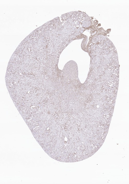

Separation of compartments in a section of mouse kidney stained for Col1a1 by immunohistochemistry. The separation is done by AI-based image analysis.
The compartments are: Blue: Cortex, Red: Outer medulla, Yellow: Inner medulla, Pink: Papilla
AI-assisted glomerular analysis
AI-assisted analysis enables in-depth evaluation of the glomerular compartments and single-cell analysis of specific stains in CKD preclinical models. This approach supports advanced digital pathology workflows for detailed cellular and subcellular analysis.
After the detection of the glomeruli is done by the AI-assisted image analysis to utilize specific immunohistochemical staining to detect glomeruli specific expression. This could be Col1a1 for fibrosis, aSMA for fibrogenesis, or Willms tumor 1 (WT1) and podocin for podocytes.
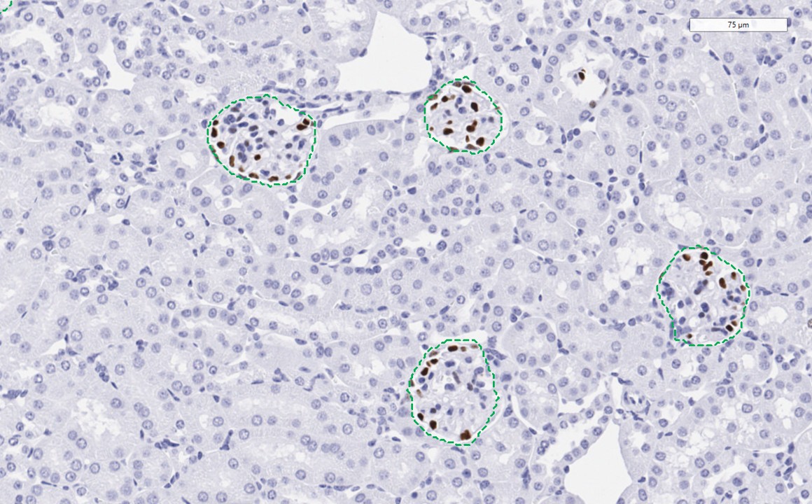
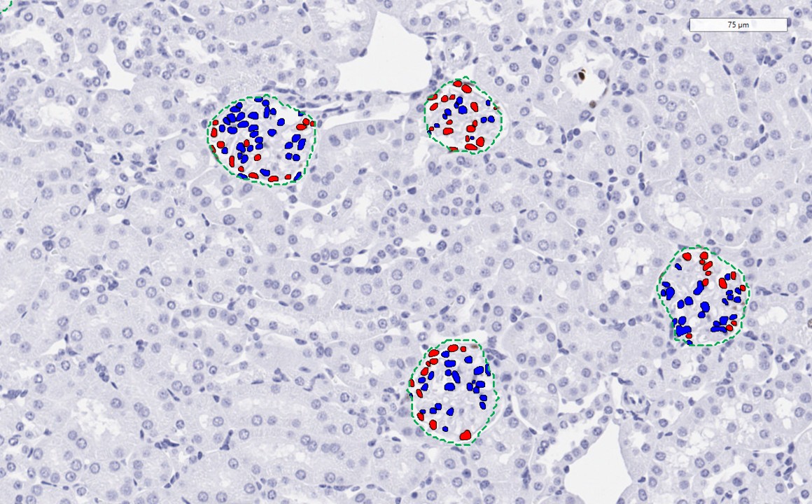
Detection of glomeruli in a murine kidney done by AI-based image analysis (left). Identification of podocytes and non-podocytes in the glomeruli (right) using an immunohistochemical stain of Willms tumor.
AI scoring and renal glomerulosclerosis histology
Glomerulosclerosis is a hallmark of chronic kidney disease (CKD) and involves the formation of glomerular scar tissue. Glomerulosclerosis refers to the progressive damage and fibrosis of the kidney’s filtering units (glomeruli), leading to a decline in kidney function.
Accurate histopathological evaluation of glomerulosclerosis is crucial for assessing the diagnosis, prognosis, and treatment of CKD patients. Several rodent models of CKD demonstrate glomerulosclerosis and are instrumental in preclinical target discovery and drug development to treat this condition, such as the Gubra UUO model.
For assessing the severity of glomerulosclerosis, glomeruli are classified from normal to complete fibrosis based on the percentage of glomerular sclerosis. This evaluation is traditionally done by a histopathologist. However, with several hundred glomeruli per sample, scoring is difficult and liable to inter-observer variability. These limitations highlight the need for automated and AI-based scoring methods in kidney pathology.
AI-based glomerulosclerosis scoring
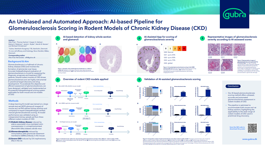
To increase objectiveness and efficient assessment of glomerulosclerosis, we have designed, validated, and implemented an AI-powered histopathological scoring system applicable for rodent models with glomerulosclerosis. Our AI-based glomerulosclerosis scoring method offers unbiased, accurate, and automated glomerulosclerosis assessment for reproducible, actionable results.
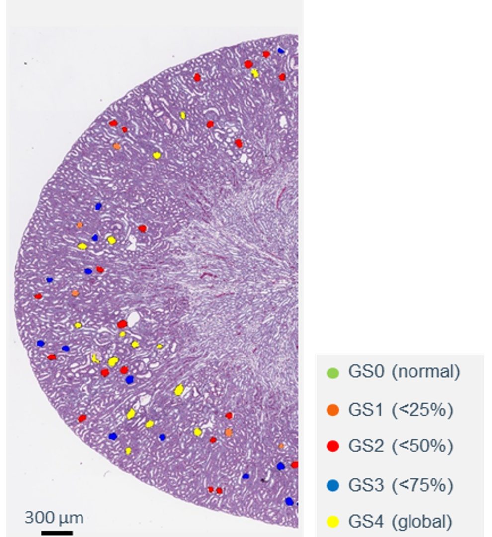
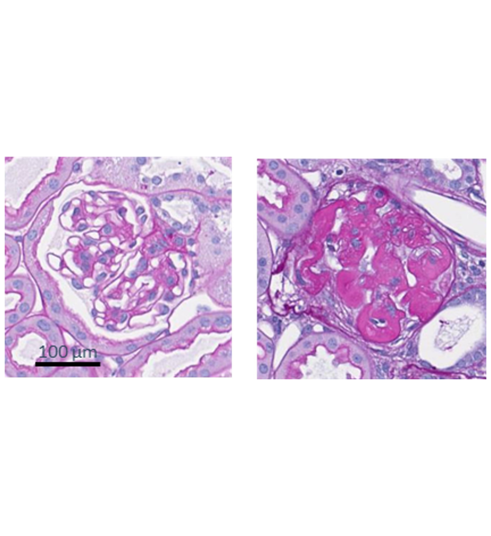
Automated detection of PAS-positive glomeruli and scoring of glomerulosclerosis by Gubra Histopathological Objective Scoring Technique (GHOST) deep learning-based image analysis. A scoring-based colour code was used to visualize sclerosis severity (GS0-GS4) in affected glomeruli.
Left panel: Representative kidney image from a vehicle-treated db/db UNx-ReninAAVmice with visualization of scoring-based color code of individual glomeruli
Right panels: Normal glomerus (left, GS0) vs. global glomerulosclerosis (right, GS4)
Animal models of kidney disease
When combined with AI-based histology and quantitative image analysis, preclinical kidney disease models enable precise, region-specific assessment of disease progression and therapeutic effects.
At Gubra, we specialize in providing high-end preclinical models tailored for your studies within chronic kidney disease (CKD), diabetic kidney disease (DKD), and acute kidney injury (AKI). Our translational models are instrumental for characterizing preclinical drug candidates and benchmarking against current treatments. We offer full-service solutions in preclinical drug development. Our pre-clinical kidney-disease models include:
| Disease | Mouse model |
|---|---|
| Chronic kidney disease | 1. Adenine-diet induced (ADI) mouse model
2. Unilateral ischemia reperfusion injury (uIRI) mouse model 3. Unilateral ureteral obstruction (UUO) mouse model 4. 5/6 nephrectomy rat model 5. Anti-glomerular basement membrane (GBM) mouse model |
| Diabetic kidney disease | Renin AAV UNx db/db mouse model |
| Acute kidney injury | Bilateral ischemia reperfusion injury (bIRI) mouse model |
These preclinical rodent models possess highly translatable phenotypes, mimicking the diverse array of human kidney disease phenotypes, such as chronic inflammation, fibrosis, impaired kidney function, and systemic issues.
To learn more about the full preclinical kidney model portfolio, explore our blog article here.
AI histology services at Gubra – Get in touch
AI-based scoring and quantitative image analysis are becoming essential tools in preclinical kidney pathology, enabling reproducible, high-throughput, and region-specific assessment of disease progression and therapeutic effects. By combining digital pathology with translational animal models, these approaches provide deeper and more reliable insights for preclinical research.
At Gubra, we integrate AI-driven histology, validated kidney disease models, and advanced biomarker analysis to support all stages of preclinical kidney research. Interested in applying AI-based histology in your study? Click here to get in touch with our team to learn more.
Nephroprotective effects of enalapril in the anti-GBM mouse model of glomerulonephritis
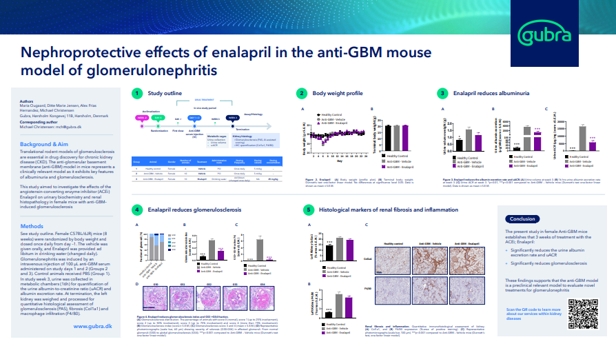
- Explore the poster here
Renal transcriptomic profiling reveals beneficial effects of semaglutide and lisinopril in advanced diabetic kidney disease

- Explore the poster here
An Unbiased and Automated Approach: AI-based Pipeline for Glomerulosclerosis Scoring in Rodent Models of Chronic Kidney Disease (CKD)
- Explore the poster here