
Amyloid plaques remain a central endpoint in Alzheimer’s disease drug development, but they are only one part of the pathology. Tau pathology, neuroinflammation, neurodegeneration and vascular changes all influence disease biology and translational interpretation. This is where quantitative 3D whole-brain imaging can add value. An Alzheimer’s disease brain scan can show amyloid or tau status in clinical research, while preclinical imaging can provide deeper regional and cellular-level resolution in Alzheimer’s mouse models. With whole-brain spatial imaging, teams can move from selected sections to atlas-aligned, brain-wide endpoints that support more confident decisions in neurodegenerative disease research.
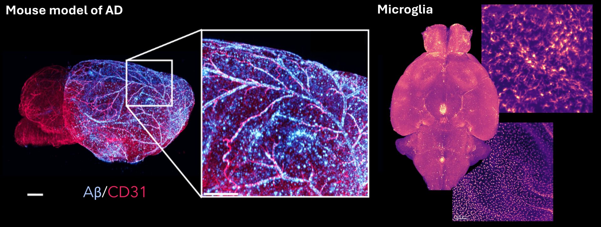
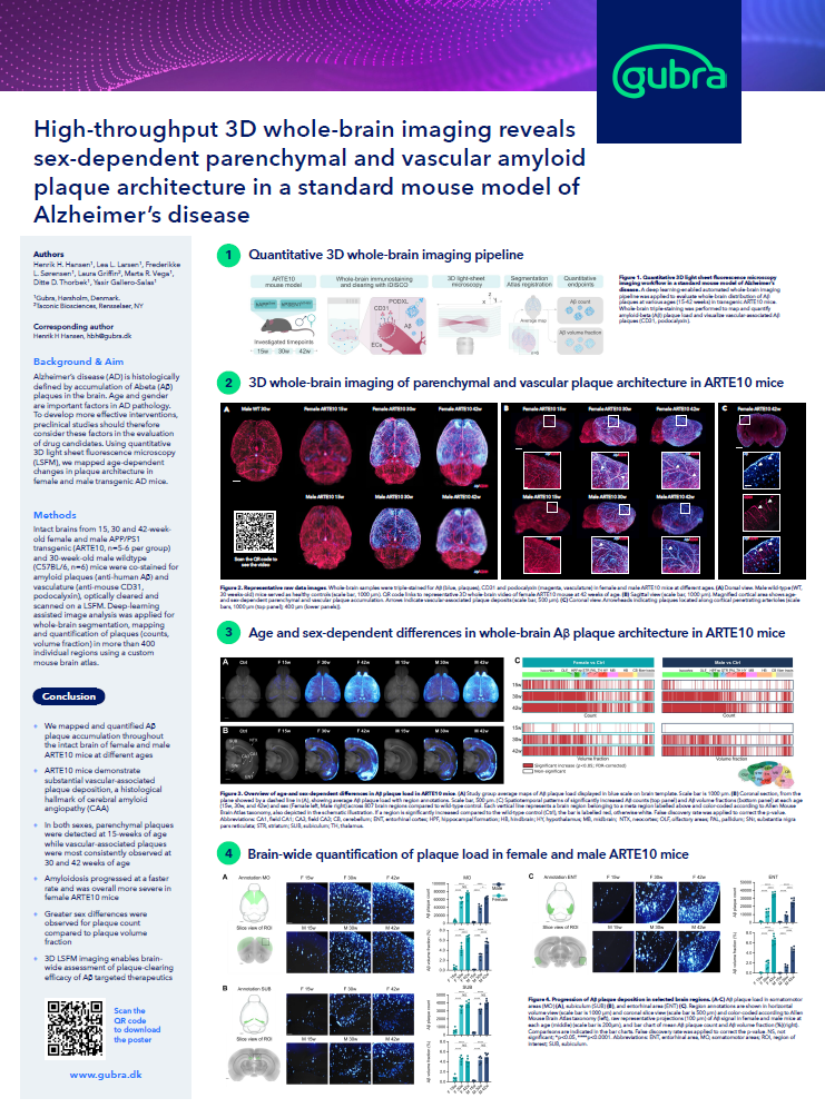
3D whole-brain imaging of amyloid pathology, vasculature and microglia in an Alzheimer’s disease mouse model
Alzheimer’s drug development is entering a new phase
Recent progress in anti-amyloid therapy has changed expectations for Alzheimer’s disease research. The discussion is increasingly about whether a treatment can engage pathology throughout the brain, reduce amyloid burden rapidly enough to be meaningful, and do so with an acceptable safety profile. At the same time, the field is moving beyond single-endpoint thinking. Amyloid reduction is important, but the therapeutic question increasingly includes whether treatment also changes tau pathology, neuroinflammatory state, neuronal stress and broader disease trajectory.
Brain shuttles have brought this question into sharper focus. A high-profile example is trontinemab, an investigational Brainshuttle™ antibody designed to improve transport across the blood-brain barrier and target aggregated amyloid. Roche has reported rapid amyloid PET changes in the Phase Ib/IIa Brainshuttle AD study, alongside changes in fluid biomarkers including phosphorylated tau. For preclinical teams, this highlights a broader point: target engagement, biomarker response and spatial tissue biology need to be interpreted together.
Why quantifying amyloid plaques, tau pathology and neuroinflammation matters in Alzheimer’s research
What is amyloid plaque?
Amyloid plaques are extracellular deposits formed mainly from aggregated amyloid beta. In Alzheimer’s disease, amyloid beta plaques are one of the defining histopathological hallmarks of the disease and a central endpoint in many Alzheimer’s disease drug development programs. However, amyloid pathology is not a single uniform signal. Plaques can differ by anatomical region, age, sex and tissue compartment. In Alzheimer’s mouse models, they may also appear as parenchymal deposits or as vascular-associated plaques, which can be relevant when evaluating cerebral amyloid angiopathy-like features and BBB-related therapeutic hypotheses.
3D whole-brain imaging of amyloid pathology and vasculature in a mouse model of Alzheimer´s Disease (AD)
What is tau pathology in Alzheimer’s disease?
Tau pathology refers to abnormal tau protein accumulation, including phosphorylated tau species and neurofibrillary tangles, closely linked to neuronal dysfunction, anatomical disease spread and cognitive decline. This makes tau an important translational readout when researchers want to understand whether a treatment changes more than amyloid burden alone.
Why does neuroinflammation matters in Alzheimer’s disease?
Neuroinflammation, caused by an increase in glial signalling and reactivity in the brain, is another key component of Alzheimer’s disease biology. Microglia respond to amyloid plaques, tau pathology, neuronal injury and vascular changes. These glial responses can be protective, harmful or context-dependent, which makes simple interpretation difficult but also makes spatial measurement highly valuable. For drug development, neuroinflammation readouts can help answer questions that plaque quantification alone cannot address:
-
- Does a treatment reduce amyloid while leaving a pro-inflammatory tissue environment unchanged?
- Does a candidate alter microglial clustering around plaques?
These questions are difficult to answer from sparse 2D sections alone, particularly in a spatially heterogeneous disease.
High-throughput 3D whole-brain imaging in Alzheimer’s mouse models
Alzheimer’s mouse models are spatially complex, and this makes whole-brain imaging particularly valuable. Gubra’s high-throughput 3D whole-brain imaging approach combines whole-brain immunostaining and clearing, light-sheet fluorescence microscopy, atlas registration and AI-assisted quantification to generate standardized endpoints from intact brains.
In the ARTE10 mouse model of Alzheimer’s disease, we, in collaboration with Taconic, used quantitative 3D light-sheet fluorescence microscopy to map age-dependent changes in plaque architecture in female and male APP/PS1 transgenic mice. Intact brains from 15-, 30- and 42-week-old ARTE10 mice were co-stained for amyloid plaques and vasculature, optically cleared, scanned, segmented and quantified across hundreds of atlas-defined brain regions.
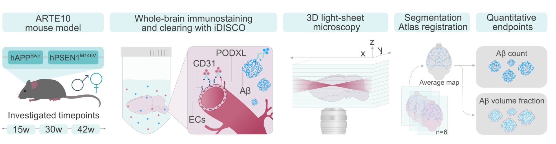
Quantitative 3D whole-brain imaging workflow, from ARTE10 model and staining to light-sheet imaging, atlas registration and quantitative endpoints
What the ARTE10 plaque imaging data show
The ARTE10 data demonstrate why spatial imaging matters in mouse models of Alzheimer’s disease. The 3D LSFM workflow enabled us to:
- Map and quantify plaque accumulation throughout intact brains in female and male ARTE10 mice.
- Detect parenchymal plaques at 15 weeks of age, while vascular-associated plaques were most consistently observed at 30 and 42 weeks.
- Identify faster and overall more severe amyloidosis progression in female ARTE10 mice.
- Assess both plaque count and plaque volume fraction.
- Support brain-wide assessment of plaque-clearing efficacy.
- Provide an amyloid and vascular endpoint example that can be paired with tau and neuroinflammation endpoints in broader AD study designs, where appropriate.
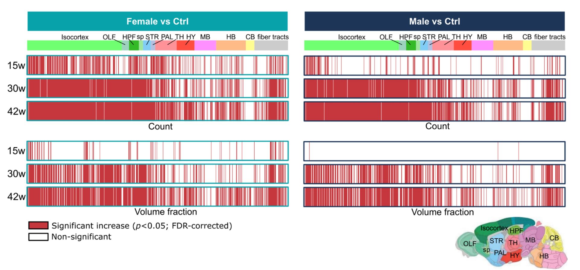
Sex- and age-dependent Aβ plaque burden in an Alzheimer’s disease mouse model
Supporting translational Alzheimer’s disease research at Gubra
For teams developing CNS therapeutics, quantitative 3D whole-brain imaging can help connect mechanism, distribution and pathology. It can complement in vivo imaging, fluid biomarkers, conventional histology and pharmacology by adding a detailed spatial endpoint across the entire intact brain.
As an Alzheimer’s disease preclinical CRO, Gubra supports translational Alzheimer’s research with disease-relevant models, whole-brain imaging, AI-assisted quantification and integrated study design. The same platform can be used more broadly in neurodegenerative disease research where spatial mapping of pathological proteins, neuroinflammation, neuronal loss or vascular remodeling is relevant.
If you are planning an Alzheimer’s disease study, exploring BBB shuttle-enhanced therapeutics, comparing plaque-clearing efficacy, or evaluating tau and neuroinflammation endpoints, quantitative 3D whole-brain imaging can provide the spatial evidence needed to make stronger preclinical decisions.
Explore quantitative whole-brain imaging for your Alzheimer’s disease program
Planning to attend AAIC 2026 in London? Meet with Gubra’s CNS and imaging experts before or during the conference to discuss how quantitative 3D whole-brain imaging can support your program.
Download Gubra’s latest Alzheimer’s disease imaging research
Our latest research demonstrates how high-throughput 3D whole-brain imaging can reveal sex-dependent differences in parenchymal and vascular amyloid plaque architecture in a standard mouse model of Alzheimer’s disease.
High-throughput 3D whole-brain imaging reveals sex-dependent parenchymal and vascular amyloid plaque architecture in a standard mouse model of Alzheimer’s disease