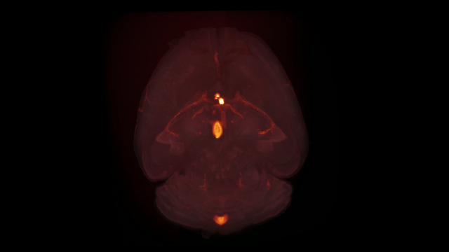

The challenge of accurately quantifying neurodegeneration in preclinical models
Neurodegenerative disease is a spatially distributed, circuit-level process rather than a focal pathology. In Alzheimer’s disease (AD) mouse models, amyloid-β (Aβ) pathology and neuroinflammation extend across cortical and hippocampal regions. In mouse models of Parkinson’s disease (PD), α-synuclein (aSyn) pathology propagates along anatomically connected pathways.
The nigrostriatal system highlights this complexity. Dopaminergic neuron cell bodies degenerate in the substantia nigra pars compacta (SNc), while their axons project to the striatum. Quantifying only tyrosine hydroxylase (TH)-positive neurons in the SNc fails to capture projection loss and striatal terminal degeneration, providing an incomplete assessment of disease burden.
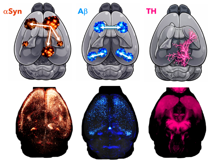
Neurodegenerative diseases involve spatially distributed pathology (αSyn, Aβ) and circuit-level cell populations (nigrostriatal TH projections)
In neurodegenerative diseases, multiple cell types shape disease progression, including neurons (NeuN), microglia (Iba1, Hexb, TREM2), astrocytes (SOX9), and blood vessels (CD31, SM22, vWF). Whole-brain staining reveals their distinct yet overlapping spatial distributions.
In preclinical drug development, accurately quantifying these endpoints is critical for robust assessment of therapeutics.
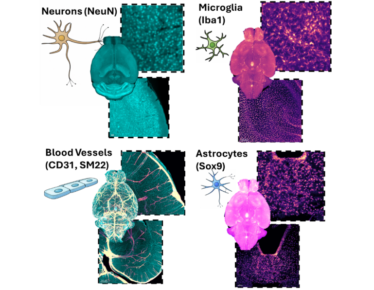
Multi-cellular landscape of neurodegeneration: neurons, microglia, astrocytes, and vasculature in whole-brain 3D imaging
Why traditional 2D histology falls short in neurodegenerative disease research
Section-based 2D histology has long been the standard in CNS research, yet it introduces inherent sampling bias that can obscure biologically meaningful effects. In neurodegenerative disease models, where pathology is spatially heterogeneous, this limitation becomes critical.
Case 1: TH+ neurons in the nigrostriatal system
In mouse models of PD, subtle dopaminergic degeneration that appears ambiguous in single sections becomes clearly detectable in volumetric reconstructions. TH terminal density changes across the striatum and neuronal loss in the substantia nigra can be assessed simultaneously resulting in greater sensitivity to early or moderate neurodegeneration.
2D Section vs 3D Volume
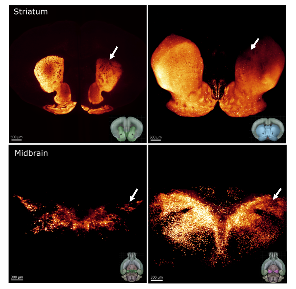
Enhanced detection of TH Soma and projection loss by 3D imaging vs. 2D sections
Case 2: Blood vessels and amyloid-β (Aβ) plaques
Amyloid-b (Ab) pathology and cerebral amyloid angiopathy (CAA) are inherently three-dimensional histopathological features of Alzheimer’s disease (AD). In traditional 2D histology, cerebral vessels appear fragmented, preventing accurate assessment of inherently 3D structures. In full volumes, however, the spatial relationship between Aβ aggregates (plaques) and the vasculature becomes evident. Notably, CAA is especially prominent in larger-calibre vessels, suggesting that vascular amyloid deposition is not spatially uniform but follows structural constraints.
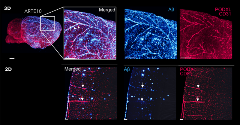
Whole-brain 3D imaging uncovers preferential Aβ deposition in large-caliber cerebral vessels
Case 3: Variability and statistical power
Physical sectioning introduces variability through cutting angle, regional sampling differences, and operator-dependent selection. Whole-brain 3D imaging eliminates sectioning bias, enabling standardized quantification across animals. Using AI-based detection algorithms for α-synuclein fibrils and Aβ plaques, regional pathology can be quantified with low inter-sample variability. In this framework, variability accurately reflects true biological differences rather than technical artifacts (see figure below, rows represent individual animals). For translational neurodegenerative disease research, reduced variability directly impacts study design:
- lower group sizes
- improved statistical power
- more robust readouts
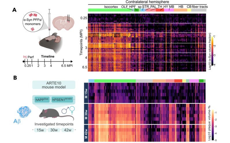
Eliminating sectioning bias: AI-driven 3D quantification reveals true biological variability
Advance your neurodegenerative research with 3D imaging
Whole-brain 3D imaging enables AI-driven quantification of pathological proteins, neuronal loss, neuroinflammation, and vascular remodelling at single-cell resolution, delivering robust, reproducible endpoints in mouse models of neurodegenerative disease. At Gubra, we have developed an end-to-end automated 3D imaging and AI driven quantification pipeline capable of imaging 500 brains per week. Gubra collaborates with pharma and biotech to design high-precision preclinical CNS imaging studies that improve sensitivity, reduce variability, and strengthen the translational value of therapeutic programs. If you are exploring preclinical strategies in neurodegenerative diseases and want to enhance the translational relevance of your CNS studies, contact our experts here.
Download our AD/PD™ 2026 posters
At AD/PD™ 2026 in Copenhagen, Gubra is presenting our latest research advancing preclinical models and translational strategies in neurodegenerative disease. Explore the featured posters and our full poster schedule at AD/PD™, add key sessions to your calendar, or connect with our team to arrange a meeting during the congress below.
Access this poster to learn how whole-brain 3D imaging captures αSyn spread and progressive TH neuron loss across the whole brain:
- Poster title: Whole-brain 3D quantification of aSyn spreading and dopaminergic neurodegeneration in a mouse model of Parkinson’s disease
- Poster number: 388
- Poster presentation: Shift 1 on the 17 – 19 March, 2026
- Explore the poster here
Download our poster to explore our scalable, AI-driven framework for automated whole-brain 3D quantification for characterization of neurodegenerative pathology:
- Poster title: AI-driven whole-brain cellular profiling using 3D microscopy
- Poster number: 1258
- Poster presentation: Shift 2 on the 19 – 21 March, 2026
- Expore the poster here
Download this poster to see how whole-brain 3D imaging enables precise, region-specific quantification of age- and sex-dependent Aβ pathology in a transgenic Alzheimer’s disease mouse model:
- Poster title: High-throughput 3D whole-brain imaging reveals sex-dependent parenchymal and vascular amyloid plaque architecture in a standard mouse model of Alzheimer’s disease
- Poster number: 1089
- Poster presentation: Shift 1 on the 17 – 19 March, 2026
- Explore the poster here
Read our publication on how TfR1-mediated BBB shuttling of Aducanumab enhances brain delivery and plaque clearance, quantified by 3D imaging in an APP/PS1 AD model:
- Publication title: Transferrin receptor-binding blood-brain barrier shuttle enhances brain delivery and efficacy of a therapeutic anti-Aβ antibody
- Expore the publication here