
Artificial intelligence is rapidly reshaping how we approach histological assessment in preclinical research, bringing greater consistency, scalability, and depth to tissue analysis. At Gubra, we operate a fully digitalized pipeline that leverages whole-slide imaging and advanced digital image analysis. In earlier articles in this series, we’ve shown how AI enables robust scoring in both:
In this article, we turn to idiopathic pulmonary fibrosis (IPF), exploring how AI-driven image analysis can enhance the evaluation of complex lung pathology and support more precise, quantitative readouts in fibrosis research.
Why is lung histology central to preclinical pulmonary disease models?
Preclinical pulmonary disease research relies on multiple evaluation endpoints, among which histological assessment of lung tissue is central. Lung histology enables detailed evaluation of fibrosis, inflammation, and general cellular injury. Evaluating the efficacy of compounds in preclinical animal disease models is an essential step before advancing to human studies, ensuring that a candidate has the potential to improve lung health. To enable translational relevance, histopathological assessment in animal models should be conducted using methodologies aligned with those applied to human lung samples.
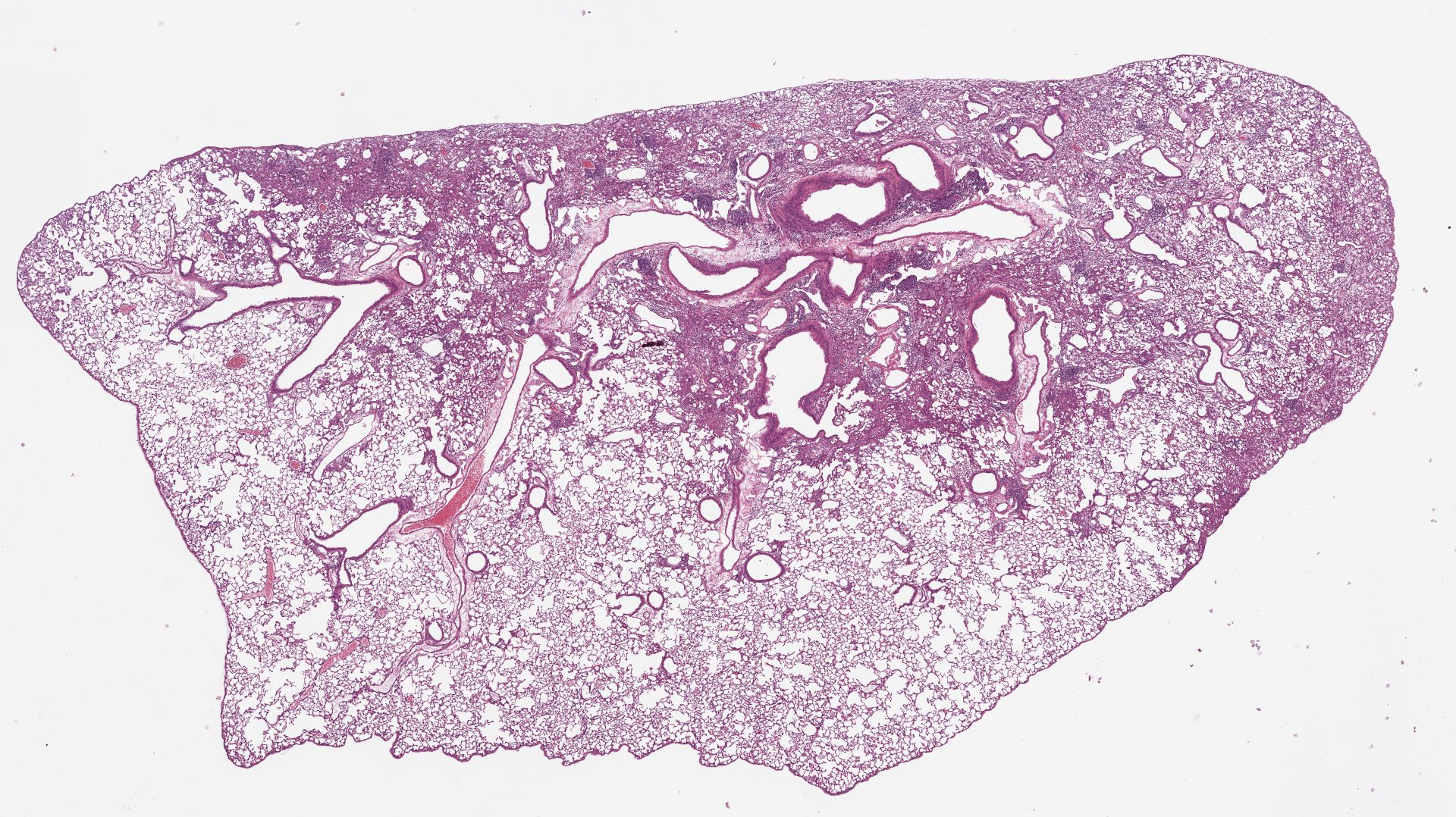
Extensive fibrosis and inflammation are found in the lung tissue seen in the areas with strong staining – stained with hematoxylin and eosin stain.
Key aspects of high-quality preclinical lung histology
Masson’s Trichrome staining forms the analytical backbone for lung fibrosis scoring, as it delineates collagen deposition and enables identification of interstitial and perivascular fibrosis. Increased blue-stained matrix within thickened alveolar septa or around bronchioles provides an immediate indicator of fibrotic remodeling and architectural distortion. We will discuss this staining more in the below section covering our AI-scoring system.
Picrosirius Red for quantitative fibrosis assessment
This fibrosis-focused staining is complemented with Picrosirius Red (PSR), to enhance detection and quantification of fibrillar collagen. PSR staining is useful for distinguishing dense, mature collagen fibers within the lung. To support robust and objective assessment, we apply advanced digital image analysis pipelines to quantify these histological features and evaluate the impact of preclinical treatments on the lung fibrosis.
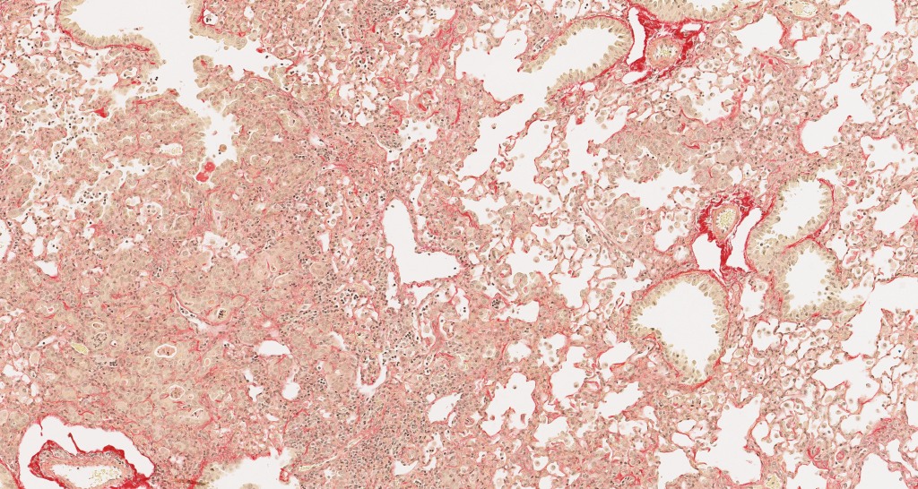
Extensive fibrosis and inflammation are found in the lung tissue noted by the strong red stain – stained with picrosirius red stain.
Immunohistochemical markers of fibrosis and senescence
At the molecular level, immunohistochemical markers such as Collagen type I (Col1a1) and Collagen type III (Col3) allow immunohistochemical assessment of extracellular matrix composition and maturation state, while α-smooth muscle actin (αSMA) labels activated myofibroblasts, a key effector population driving fibrogenesis. To evaluate tissue injury and remodeling dynamics, we assess p21 as a marker of cellular senescence and cell-cycle arrest, and Galectin-3 (Gal-3) as an indicator of macrophage activation and pro-fibrotic signaling. At Gubra, we offer customized histochemical and immunohistochemical staining panels tailored to specific preclinical animal study endpoints in mouse and rat models. All markers are quantified using digital image analysis for clear and reproduceable readouts.
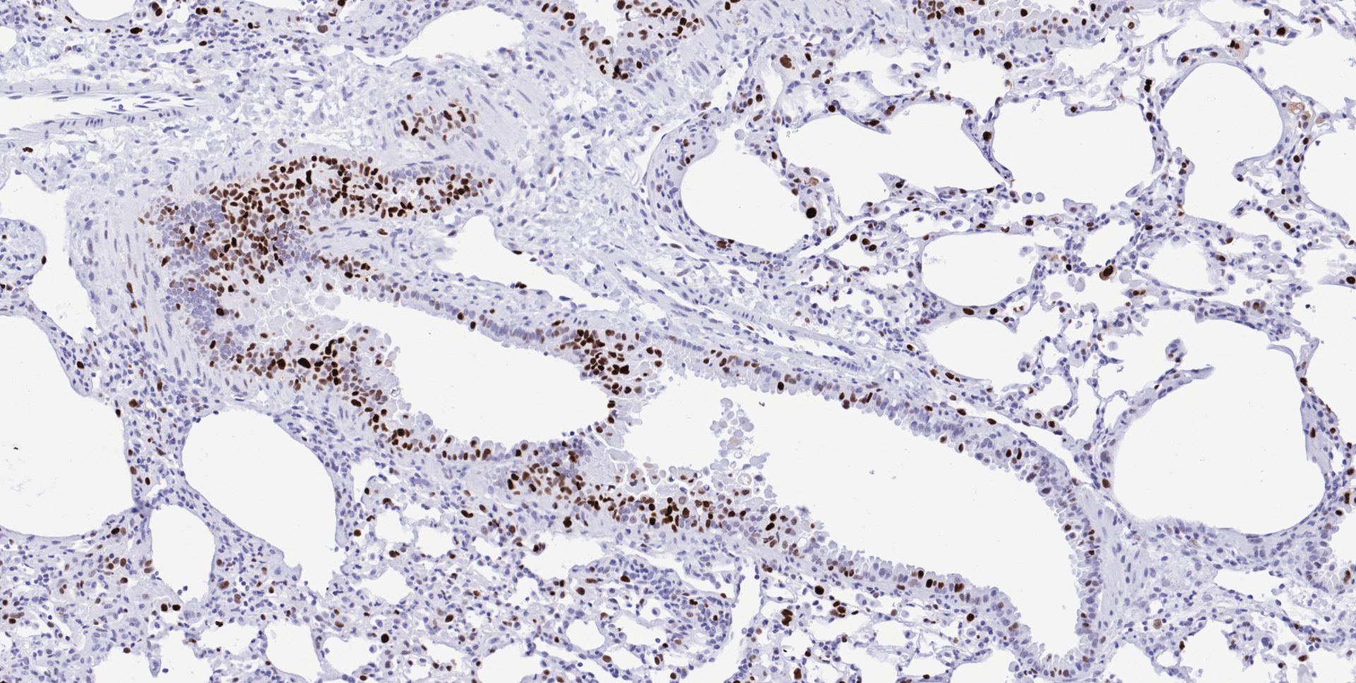
Bronchi with epithelium positive for p21 demonstrating that these are senescent cells – stained in brown.
AI-based histological evaluation of fibrosis in preclinical lung models ensures reproducibility
In preclinical models for Idiopathic Pulmonary Fibrosis (IPF) and other fibrotic pulmonary conditions, the Ashcroft scoring system is widely used for the histological assessment of fibrosis in the pulmonary tissue. This scoring system was originally developed to evaluate pulmonary fibrosis in clinical settings and is now applied to preclinical rodent models to draw parallels between efficacy in animal models and the treated patients. The Ashcroft scoring system has nine different classes, from 0 to 8, classifying fibrotic patterns in the lung tissue stained with Masson’s Trichrome to delineate collagen deposition.
Like other manual, semiquantitative histopathological methods, Ashcroft scoring system is subject to inter- and intra-observer variability, affecting the accuracy and reproducibility of study outcomes. To remove this variability in preclinical studies, at Gubra we have developed an automated, deep learning-assisted digital image analysis method, termed GHOST (Gubra Histopathological Objective Scoring Technology), for objective assessment of Ashcroft scores in a spirometry-confirmed, bleomycin-induced idiopathic pulmonary fibrosis mouse model.
The model identifies the severity of fibrosis via intensity of the Masson’s Trichrome staining in tiles digitally segmented onto the lung section (see below). A tile with healthy looking tissue will receive a score of 0, while severely fibrosed tissue will receive a score of 8. Tiles predominantly occupied by large bronchi or vessels are ignored. These scores are then averaged for an individual and eventually group wide score for highly unbiased and precise evaluation of hundreds of samples at a time.
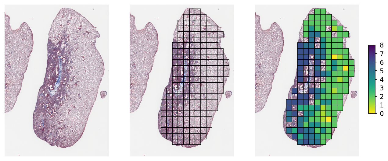
Automated deep learning-based Ashcroft scoring of lung fibrosis in mouse with IPF.
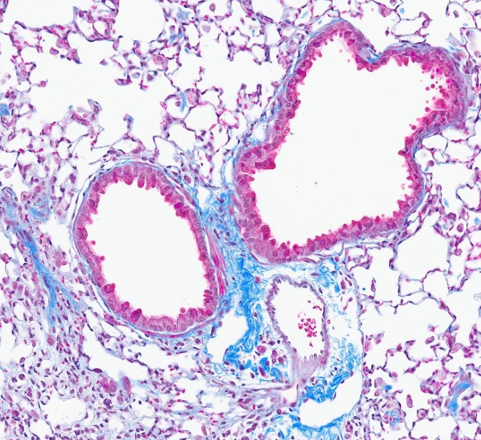
Lung tissue from a mouse with bleomycin-induced fibrosis – Masson’s Trichrome stain.
AI histology services at Gubra – Get in touch
See the posters below to dive deeper into how we utilize our AI-based, Ashcroft scoring system to evaluate the efficacy of compounds used in our IPF mouse model. Interested in using this scoring system? Reach out to learn more about AI‑based histological quantification at Gubra.
Automated AI-assisted Ashcroft scoring of lung fibrosis in spirometry-confirmed and bleomycin-induced mouse model of IPF
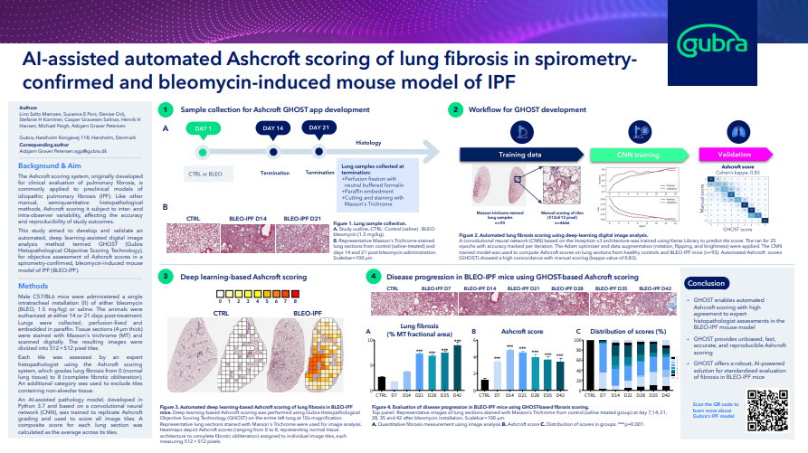
- Explore the poster here
Molecular Hallmarks of Lung Cellular Senescence in the Repetitive Bleomycin-induced and Spirometry-confirmed Mouse Model of IPF
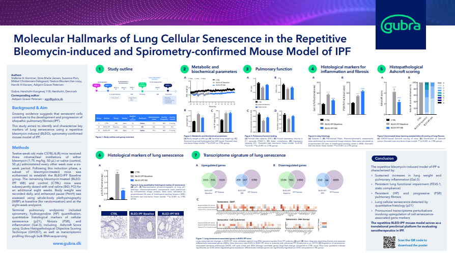
- Explore the poster here
Reproducible lung protective effects of a TGFβR1/ALK5 inhibitor in a bleomycin-induced and spirometry-confirmedmodel of IPF in male mice
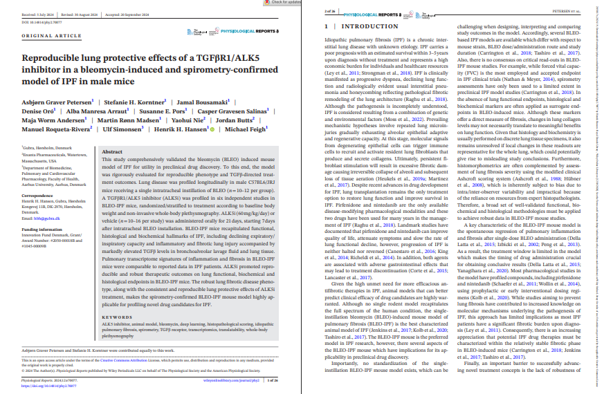
- Explore the poster here