
What are preclinical histology services in drug discovery?
The process of drug discovery from target identification through clinical trials is a lengthy process that requires clear and consistent data to evaluate both efficacy and safety. During the preclinical phase of drug discovery, histology is one of the technologies employed to evaluate the potential impact. The histological endpoints can, among others, include changes to tissue morphology, characterization of cellular reaction patterns, altered function, protein or gene expression, tumor burden, or compound distribution. Histology read-outs on tissue from preclinical animal models evaluate the translation of a therapy and frame the results in a human-specific context. This type of preclinical histology work can greatly accelerate drug development and IND filings for final FDA approval. At Gubra, we utilize evaluation of histological samples to deliver translational and reproducible animal models of MASH, obesity, and other metabolic and fibrotic diseases.

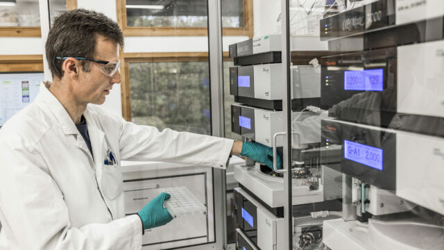
Sectioning of paraffin embedded samples for further processing
Key aspects of high-quality preclinical histology
For reproducible, high-quality preclinical histology one must:
- Ensure correct and timely handling and processing of the tissue samples starting from the time of sampling
- Use precise sectioning to correctly for identification of representative areas of interest
- Employ the correct histological staining technique, whether that be a histochemical stain like H&E, use of an antibody for immunohistochemical techniques, or RNA probe for in situ hybridization
- Utilize standardized, unbiased scoring techniques and image analysis to assess the tissue in a manner that can be repeated between batches and studies
In addition to these best practices, histology is an ever-evolving field where new approaches, such as large multiplexing experimental designs or spatial transcriptomics, allow for an ever-growing arsenal of techniques to evaluate preclinical samples.
Liver histology performed in preclinical studies for liver diseases at Gubra
Translational preclinical animal models for liver disease, such as MASH (metabolic dysfunction-associated steatohepatitis), are highly similar to human disease. At Gubra, we evaluate pre-treatment biopsies in our MASH mice to determine changes in the liver according to NAFLD-score and fibrosis stage following diet induction of MASH. This enables our customers to acquire data on each individual animal’s baseline; then, at the completion of a study, we compare the pre-treatment samples to the termination samples to establish treatment response within each individual animal and across cohorts. Additionally, we assess end points of inflammation, collagen deposition, and alternative fibrosis markers for a full picture of liver health.
Liver histology is also used to evaluate liver health in preclinical rodent models of aging, obesity, and other metabolic diseases. During aging, similar fibrosis and inflammation are observed in the liver as seen in diet-induced models of MASH or obesity. Preclinical studies hinge on the liver histology performed being consistent and unbiased to best evaluate a compound’s efficacy.
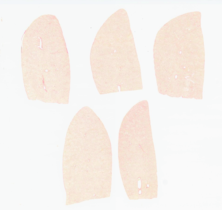
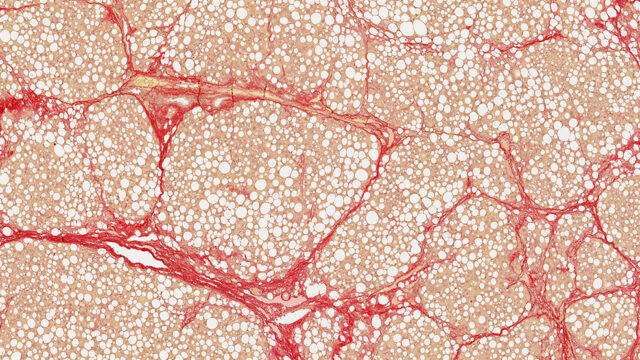
Liver biopsies from mice fed with GAN-DIO MASH diet – stained with Picro-sirius Red
From traditional liver histology to AI-driven quantification
While histology and histopathology are century-old techniques, AI and other automation techniques have greatly improved the quality and quantity of histopathological samples that can be employed in preclinical studies.
Histopathological scoring is a qualitative assessment of the morphological changes in the tissue set by re-defined criteria. However, manual histopathological scoring systems are prone to inter- and intra-observer variability which can influence robustness and reproducibility of study results. Drug efficacy studies in animal models of metabolic dysfunction-associated steatohepatitis (MASH) typically use the clinical-derived NAFLD Activity Scoring (NAS) and fibrosis staging system, outlined by Kleiner et al (2005).
Histopathological scoring in MASH
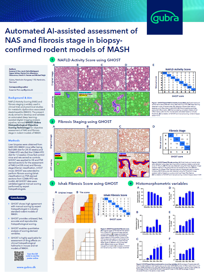
The NAFLD Activity Score is a widely used clinical scoring system for assessing the severity of non-alcoholic fatty liver disease. The NAFLD Activity Score (NAS) is a 0-8-point scoring metric comprising liver steatosis (score 0-3), lobular inflammation (0-3), and hepatocyte ballooning (0-2). These features are all assessed in a hematoxylin eosin stain (H&E) and are evaluated by morphological features of the tissue. Tissue given a high score is more severely affected than in tissue with a low score. In parallel the fibrosis staging system includes morphological features including localization and appearance of the fibrosis that graduates from no liver fibrosis (score 0) to cirrhosis (score 4). This score can be done on any histochemical stain for fibrosis and is traditionally done using Picrosirius Red.
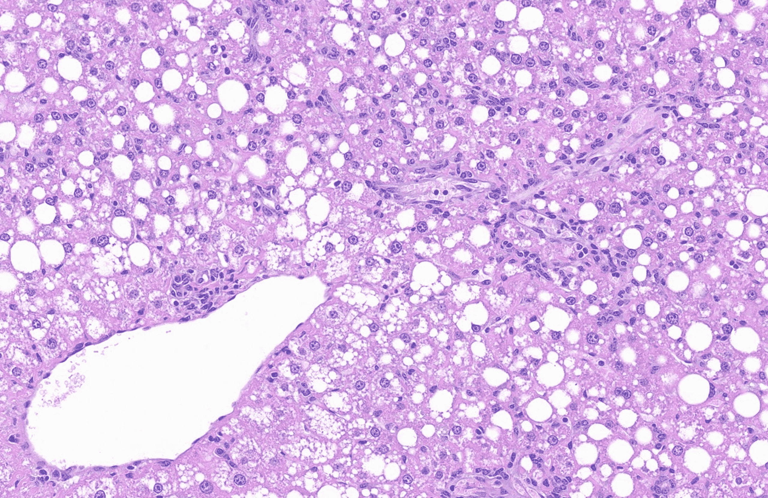
Liver from mice demonstrating liver steatotis and inflammation – stained with hematoxylin eosin stain
Gubra Histopathological Objective Scoring Technique (GHOST)
To enable objective and unbiased histopathological assessment in liver biopsies from mouse models of MASH, we developed Gubra Histopathological Objective Scoring Technique (GHOST), an automated deep learning-based digital imaging analysis pipeline for the NAS and fibrosis staging system.
To build the analysis pipeline, liver biopsies were obtained from the models of GAN-DIO-MASH done at Gubra and stained with H&E for NAS scoring and Picrosirius Red for fibrosis stage scoring. The AI model was then trained by receiving a large amount of data consisting of images scored made by histopathologists. After learning from this data, automated GHOST deep learning computational analysis were then validated against manual scoring by an expert histopathologist.
These preclinical mouse liver histology AI-scoring systems allow us at Gubra to provide rigorous, translational data from our preclinical mouse models.
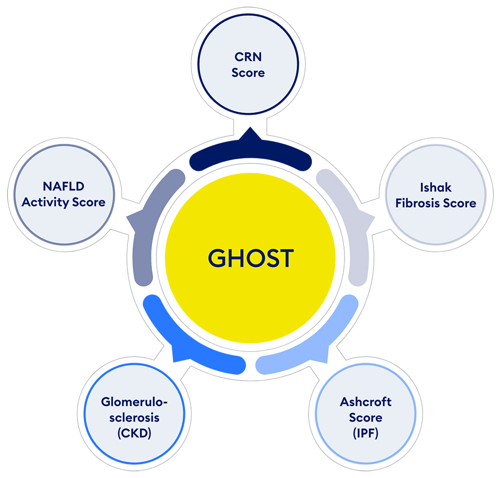
Gubra Histopathological Objective Scoring Technique (GHOST)
AI histology services at Gubra – Get in touch
If you are interested in learning more about how we use AI in histology at Gubra, contact us, follow this blog series, or read more in select posters below.
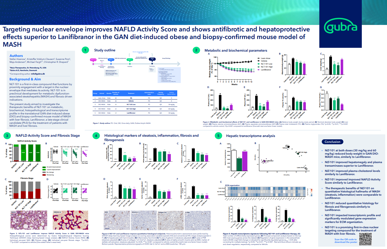
See our work using GHOST to score the effects of Lanifibranor in our MASH mouse model. Explore the poster here.
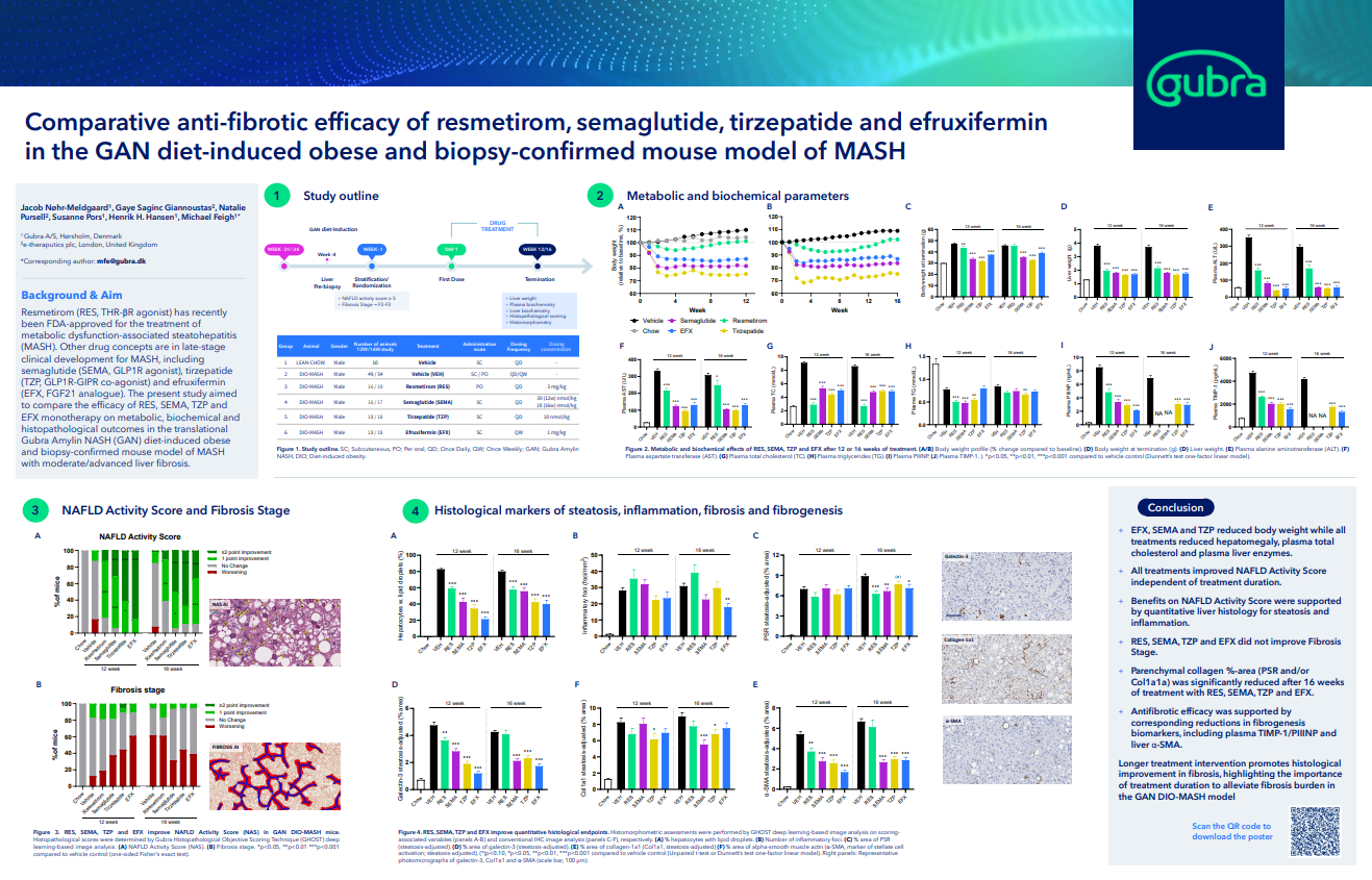
Compare treatments for MASH scored using our AI-based scoring system. Explore the poster here.
Learn more about our GHOST scoring system and how we developed our MASH scoring protocol. Explore the poster here.