

What is the blood-brain barrier?
The blood-brain barrier (BBB) is a highly selective interface between the circulating blood and the central nervous system, a specialized biological barrier that helps control which substances can enter the brain from the bloodstream.
This barrier is essential for normal brain function. The brain depends on a tightly regulated environment, where nutrients, ions, signaling molecules, and waste products are carefully controlled. At the same time, the brain must be protected from toxins, pathogens, inflammatory factors, and other circulating substances that could disrupt neuronal activity.
Blood-brain barrier structure and function
The blood-brain barrier is formed mainly by specialized endothelial cells lining the brain’s microvessels. These cells are connected by tight junctions, which restrict passive movement between cells. The barrier is further supported by pericytes and astrocytic endfeet.
The core blood brain barrier function is to maintain CNS homeostasis. It regulates the movement of molecules between blood and brain tissue, supports nutrient supply, removes waste products, and helps protect neural circuits from harmful systemic influences.
Selective permeability and transport mechanisms
The blood-brain barrier is selectively permeable. Some small lipophilic molecules can diffuse across the barrier, while essential nutrients such as glucose and amino acids enter through dedicated transport systems, for instance receptor-mediated transcytosis. These pathways are especially relevant in CNS drug delivery, where researchers aim to improve brain exposure without disrupting the protective role of the barrier.

In BBB capillaries, endothelial tight junctions regulate blood–brain exchange and are supported by surrounding cells, including pericytes, v astrocytes, perivascular OPCs, interneurons, macrophages, and microglia.
The blood-brain barrier in drug development
For CNS therapeutics, the blood-brain barrier represents a technical challenge. Its protective function helps maintain brain homeostasis, but the same selective barrier can prevent therapeutic compounds from reaching disease-relevant targets in the central nervous system.
This makes the BBB a key consideration throughout CNS drug development, from early molecule design to preclinical studies of brain exposure, target engagement, and therapeutic efficacy. A compound may show strong activity in vitro, but still fail to produce meaningful effects in vivo if the compound cannot cross the blood-brain barrier, remains trapped in the vasculature, or is actively transported back into the bloodstream.
Challenges in CNS drug delivery
One of the main drug delivery challenges in CNS research is achieving sufficient brain exposure without compromising the protective function of the BBB. Many therapeutic modalities, including antibodies, peptides, enzymes, and other large molecules, have limited natural access to the brain.
Even small molecules can face barriers such as poor permeability, rapid clearance, plasma protein binding, or active efflux. As a result, CNS drug delivery is not only about whether a compound enters the brain, but also where it distributes, how long it remains there, and whether it reaches the intended cellular or pathological target.
This is especially relevant in neurodegenerative diseases, where targets such as amyloid beta plaques, inflammatory cell populations, or vulnerable neuronal circuits may be unevenly distributed across brain regions. Preclinical studies that combine disease-relevant models with spatial readouts of drug distribution and pathology can therefore provide important insight into whether a therapeutic strategy is likely to translate into meaningful CNS efficacy.
How blood-brain barrier preclinical research services support CNS drug discovery
For BBB-focused therapeutics, the key question is not only whether a molecule can cross the barrier, but whether it reaches the right brain regions, engages the intended target, and produces a measurable biological effect.
In vitro and in vivo BBB models
In vitro BBB models are often used early in drug development. These systems can provide useful initial information however, they do not fully reproduce the complexity of the intact blood-brain barrier. Many models struggle to represent true BBB tightness, show high variability between systems, and may have limited translational value when predicting brain exposure in vivo. For this reason, in vivo preclinical studies remain essential for understanding how a therapeutic behaves in the brain. Disease-relevant animal models can help assess whether a compound crosses the BBB, where it accumulates, and whether it reaches pathological targets such as amyloid beta plaques.
TfR1-shuttled anti-Aβ antibody biodistribution across the mouse brain 24 hours after intravenous dosing.
Data generated in collaboration with Taconic Biosciences.
Advanced 3D Imaging
3D imaging adds spatial context to BBB research by showing not only how much therapeutic signal is present, but also where that signal is located. Instead of relying only on bulk measurements or selected 2D tissue sections, whole-brain 3D imaging provides a more complete view of compound distribution across relevant CNS regions and disease pathology.
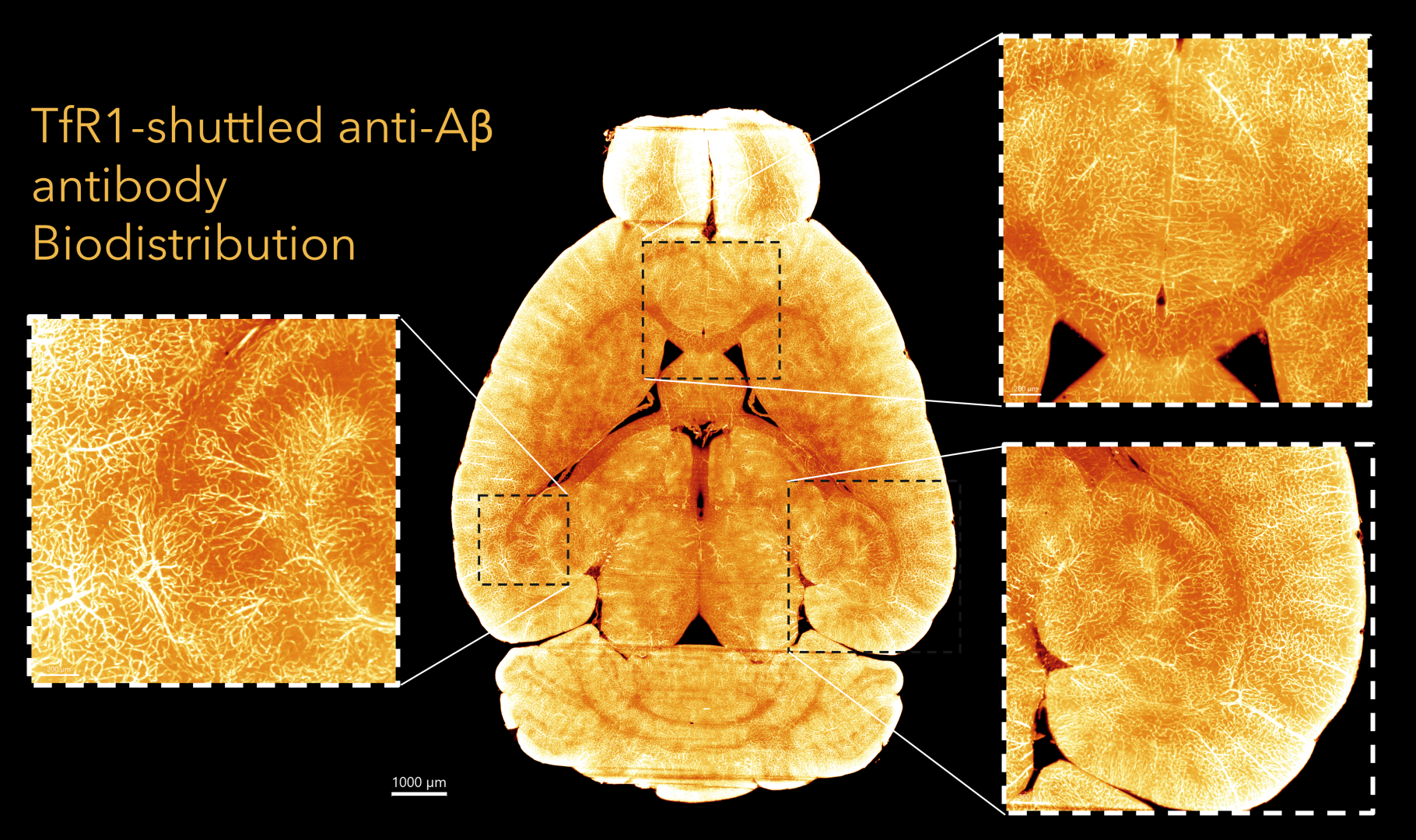
Horizontal plane view of TfR1-shuttled anti-Aβ antibody biodistribution with high-magnification regions across the hippocampus, cortex, and lateral septum.
This is particularly valuable when evaluating BBB shuttle technologies, antibody delivery, oligonucleotide biodistribution, or treatments targeting unevenly distributed pathology. For example, in neurodegenerative disease models, Gubra can image therapeutic distribution alongside pathological readouts such as beta amyloid plaques, allowing researchers to assess brain exposure and disease biology in the same brain with spatial context.
In BBB-focused studies, Gubra’s 3D imaging platform can also be combined with vascular co-labeling. For example, we use CD31 to visualize and segment the brain vasculature for quantitative analysis of therapeutic signal in relation to blood vessels, helping distinguish vessel-associated signal from true parenchymal distribution. By measuring signal inside or close to the vascular compartment versus signal outside the vessels, researchers can better assess whether a compound remains confined to the bloodstream or successfully crosses the BBB and distributes into brain tissue.
CD31 co-staining combined with therapeutic signal detection enables quantification of signal within vascular and parenchymal compartments.
Data generated in collaboration with Taconic Biosciences.
By combining tissue clearing, light-sheet fluorescence microscopy, vascular co-labeling, and quantitative image analysis, Gubra supports preclinical BBB studies that integrate CNS drug delivery with spatial characterization of disease biology. These approaches can provide deeper insight into brain penetration, target engagement, and regional therapeutic localization during CNS drug development.
Horizontal flythrough of TfR1-shuttled anti-Aβ antibody biodistribution across the mouse brain, moving from anterior to posterior regions.
Data generated in collaboration with Taconic Biosciences.
Blood-brain barrier research at Gubra – Get in touch
The materials below highlight examples of how BBB shuttle technologies and whole-brain 3D imaging approaches can support the development of CNS therapeutics with improved brain delivery and spatial target engagement.
Comparative quantitative 3D whole-brain mapping of Tfr1-enabled and unmodified anti-β-amyloid antibody biodistribution
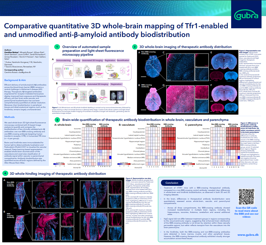
- Explore the poster here
High-throughput 3D whole-brain imaging reveals sex-dependent parenchymal and vascular amyloid plaque architecture in a standard mouse model of Alzheimer’s disease
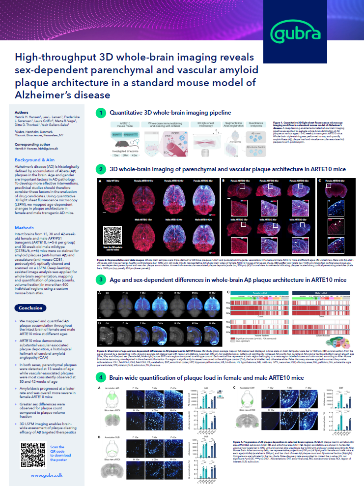
- Explore the poster here
Transferrin receptor-binding blood-brain barrier shuttle enhances brain delivery and efficacy of a therapeutic anti-Aβ antibody
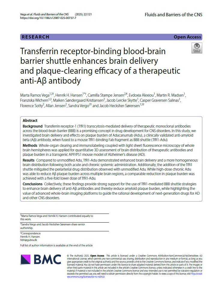
- Explore the poster here
Transferrin receptor-binding blood-brain barrier shuttle enhances brain delivery and efficacy of a therapeutic anti-Aβ antibody
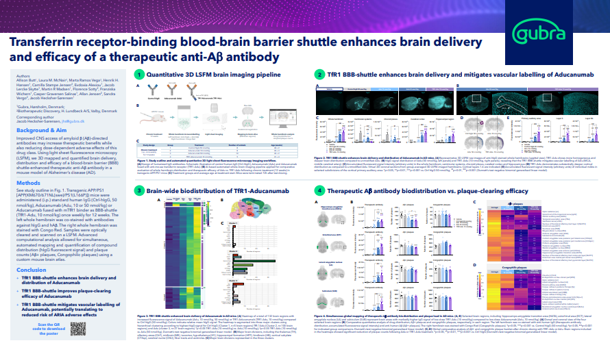
- Explore the poster here


