SCIENCE OF CERTAINTY
Simplifying the complexity of neuroscience using quantitative whole brain histology
Book a meeting with us
Gubra helps companies visualize drug distribution in the whole brain and whole organs at single cell resolutions, as well as precisely quantify histological markers. Book a meeting with us and explore how Gubra can accelerate your drug development.
Complete preclinical solutions including pharmacology, whole brain imaging and quantitative analyses
![2022-03-18_Figures_for_FoN_paper_PPT[1]](https://www.gubra.dk/wp-content/uploads/2023/04/2022-03-18_Figures_for_FoN_paper_PPT1.jpg)
High throughput imaging and analysis
- Perform IND or NDA supporting studies of up to 100 mouse brains in one study
- Fully automated whole organ immunolabelling
- AI assisted algorithms for brain mapping and quantification
- Approximately 2.5 months from study to data presentation
Our in house developed brain atlases and GubraView is the foundation of our CNS platform
![figure_gubraview2[1]](https://www.gubra.dk/wp-content/uploads/2023/04/figure_gubraview21.png)
Using reference brains to coordinate surgery and reporting of data
- Use precise in-skull coordinates for high precision stereotactic surgery
- Bridge between different atlas spaces
- Create digital brain maps with anatomical annotation
- Use GubraView to interact with your data using our digital brain browser
Optimized Mouse Brain Atlas for Automated Mapping & Quantification of Neuronal Activity
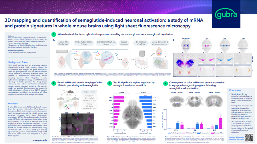
Use whole brain c-Fos imaging to better understand how drugs work in the CNS
Whole-brain Activation Signatures of Weightlowering Drugs
Unprecedented resolution and sensitivity allows gene expression to be detected in whole mouse brains
Now it is possible to detect mRNA in intact organs with unprecedented sensitivity.
Using whole brain in situ hybridization it is possible to multiplex up to three genes simultaneously. This can be used to map the expression of druggable receptors or other genes of interest. In combination with gene therapy it can be used to detect payloads such as mRNA or ASO’s. The digital maps produced are compatible with c-Fos maps or NeuroPedia maps.
![images_sec5[1]](https://www.gubra.dk/wp-content/uploads/2023/04/images_sec51.png)
Detecting mRNA in Intact Organs with Incredible Sensitivity
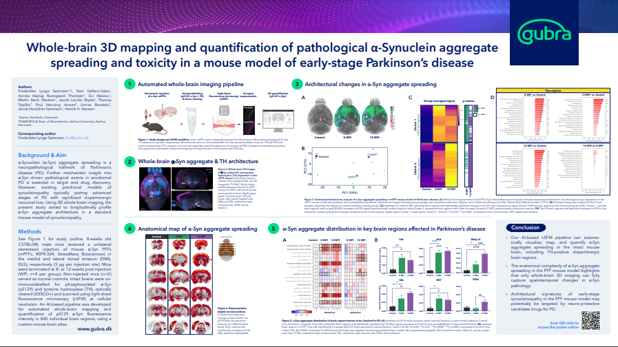
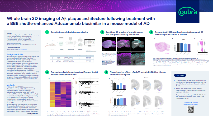
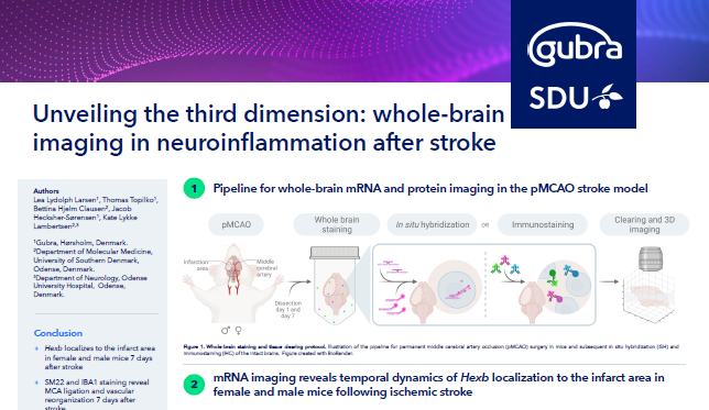
Quantify whole-brain histopathological hallmarks in mouse models of neurodegenerative diseases
Use 3D imaging to quantify multiple neurodegenerative endpoints such as plaques, dopaminergic cells and spreading of pathologies such as a-Synuclein or p-Tau.
![images_sec6[1]](https://www.gubra.dk/wp-content/uploads/2023/04/images_sec61.jpg)
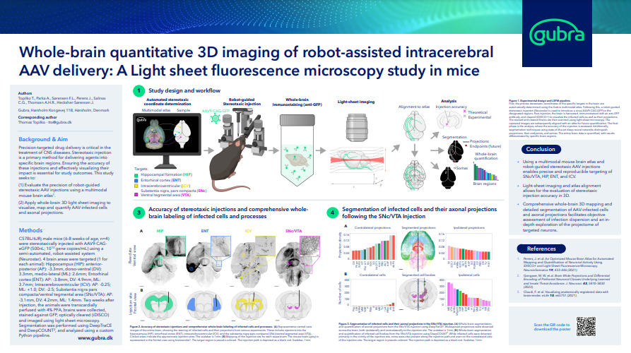
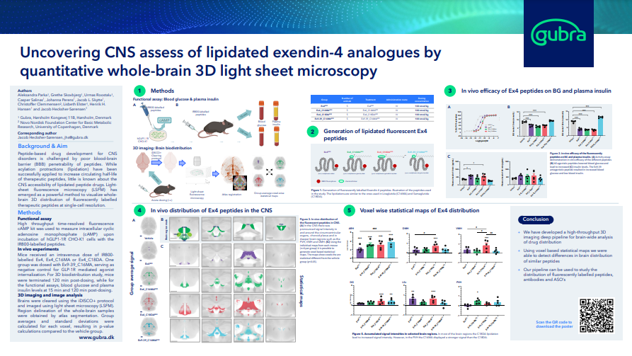
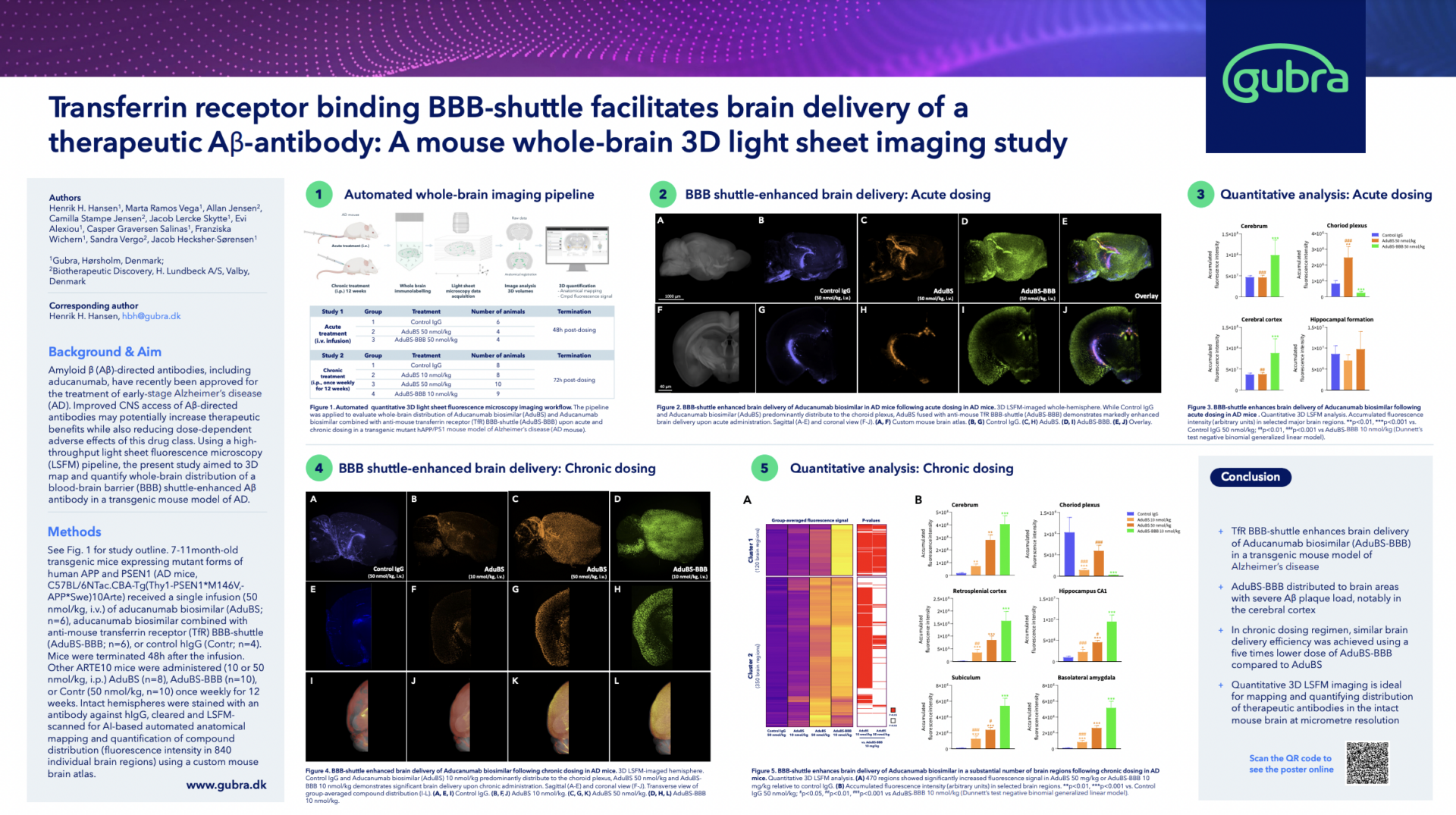
Whole brain imaging is the perfect tool to study gene therapy and biodistribution of CNS-targeted drugs
3D imaging can be used to quantify and visualize where in the brain a drug is ending up. The technology can be applied to both fluorescently labelled antibodies and peptides. For gene therapy it is possible to visualize payloads delivered using both viral and non-viral approaches. In addition it is possible to combine conventional 2D histology with 3D imaging. After imaging the brains can be sectioned and stained with additional antibodies.
![images_sec7[1]](https://www.gubra.dk/wp-content/uploads/2023/04/images_sec71.png)
Explore our data
Download the Latest Posters
Download our latest posters and explore how Gubra utilizes advanced 3D whole-brain imaging to help progress your drug discovery and development.

Jacob Hecksher-Sørensen is Director of the imaging facility at Gubra, and he has worked for more than 20 years with immunolabelling, tissue clearing and 3D imaging. Initially the technology was primarily applied to mouse embryos but over time methods and equipment have evolved to a point where it is possible to label and image adult mouse organs at single cell resolution. Today we visualise diseases progress and translate these finding into new therapies.
