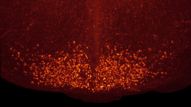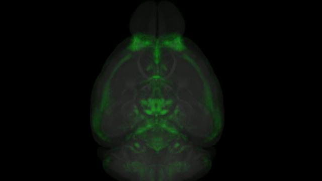

Understanding how the central nervous system (CNS)—the brain and spinal cord—drives behaviour and disease is a key focus in neuroscience. To do this, researchers often need to measure neuronal activation in response to different stimuli or compounds. A widely used tool for this purpose is c-Fos, a protein whose expression can be visualized at the single-cell level throughout the brain.
In this article, we dive deeper into c-Fos, its function, and how it can be used in research.
- For more on c-Fos and how it could augment your studies, see our blog post here: Understanding c-Fos and its applications in CNS research
What cells express c-Fos?
c-Fos is expressed in a wide range of human tissues, as seen in the Human Protein Atlas. Within the CNS, c-Fos expression is found in primarily in neurons but is expressed in glia under certain conditions.
Interestingly, c-Fos is classified as a proto-oncogene, meaning its mutation or misregulation can result in tumour formation. However, Mahner, et al., observed that in ovarian cancer higher expression of c-Fos correlates with increased survival rate. Beyond ovarian cancer, c-Fos levels have been studied in tumours in ovary, bone, endometrium, cervix, breast, intestine, lung, thyroid, and liver, as described in Durchdewald, et al.
What is the function of the c-Fos protein?
c-Fos plays a key role in helping cells, especially neurons, respond dynamically to external and internal stimuli. When a neuron is activated and a calcium influx occurs, a cascade of intracellular signalling leads to rapid transcription of the c-fos gene. Lara Aparicio, et al., summarize the c-Fos activation pattern following stimulation as follows:
- 5-20 minutes: the c-fos gene is transcribed
- Within 30 minutes: c-fos transcript levels peak
- 20-90 minutes: the c-Fos protein is translated
- 2-5 hours: c-Fos persists (half-life)
After translation, the c-Fos protein is transported to the nucleus where it becomes a part of the AP-1 transcription factor complex. This complex activates genes that enable the neuron to respond rapidly and appropriately to the initial stimulus.
Of note, c-Fos also participates in its own negative feedback loop as a member of the AP-1 complex to silence its own gene productions. This, coupled with its relatively short half-life, ensures that c-Fos expression is transient—enabling neurons to be responsive to new inputs while avoiding prolonged activation and aberrant signalling.
What is c-Fos a marker for? What does c-Fos measure?
In research, c-Fos is used as a proxy marker for neuronal activation. Using anti-c-Fos antibodies, scientists can detect and quantify the presence of c-Fos in their sectioned or whole brain samples. A c-Fos signal indicates that these neurons were activated (either directly or indirectly) by a given stimulus, such as a drug or behavioural task.
Immunohistochemistry for c-Fos offers a snapshot of the brain’s activation state at the time of collections. More expression of c-Fos typical equates to more neuronal activity. Using either immunofluorescence or chromogenic staining, researchers can visualize and quantify this activity in specific brain areas.
Work with Gubra to advance your research with c-Fos mapping
c-Fos is a useful marker that enables researchers to visualize neuronal activation with high spatial resolution, especially in whole brain imaging. Its dynamic expression in response to stimuli makes it ideal for studying how the brain reacts to behavioral, pharmacological, or environmental inputs.
At Gubra, we perform whole brain c-Fos imaging to provide you with detailed data on your compound’s effects, allowing you to identify mechanisms of action, drive new hypotheses, or differentiate between lead candidates. Contact us to learn more about how we can support your research with c-Fos imaging and analysis here.