
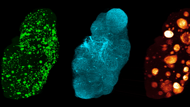
Why female models matter in preclinical research
High-quality preclinical women’s health research depends on animal models that reflect female biology. Ovarian function, reproductive hormone signaling, metabolism, and the estrous cycle can influence disease phenotypes and treatment responses. These factors are relevant not only to reproductive conditions such as polycystic ovary syndrome (PCOS), primary ovarian insufficiency (POI), and menopause, but also to systemic diseases such as diabetic cardiorenal disease.
Our previous article, Closing the preclinical gap: How better research models can transform women’s health, explored the broader need for greater female representation in research. Here, we focus on the practical application of women’s health animal models, including complementary mouse and rat models of PMOS/PCOS and menopause, as well as a female ReninAAV UNx db/db mouse model of cardiorenal disease. Together, these models allow researchers to select the disease biology, species, and endpoints that best align with their therapeutic mechanism and stage of drug development.
Preclinical models for ovarian health and function
Which features should a translational PMOS/PCOS mouse model reproduce?
Polycystic ovary syndrome (PCOS), recently renamed polyendocrine metabolic ovarian syndrome (PMOS), is not defined by a single phenotype. The updated name better reflects the interconnected metabolic, endocrine, and ovarian dysfunction that characterizes the condition. A translational PMOS preclinical model should therefore reproduce treatment-relevant biology across all three domains:
- Metabolic dysfunction: changes in body composition, insulin levels, and glycemic control
- Endocrine dysregulation: androgen excess or altered sex-hormone signaling
- Ovarian dysfunction: disrupted estrous cycling, impaired follicle development, follicular arrest, and anovulation
PMOS occurs across a range of body compositions, making it important to represent both obesity-associated and lean disease phenotypes in preclinical research. Gubra’s complementary mouse and rat models capture this heterogeneity, allowing researchers to select the model that best matches a therapeutic candidate’s mechanism of action, endpoints, and intended patient population.
DHT and Western diet-induced mouse model: Obesity-associated PMOS
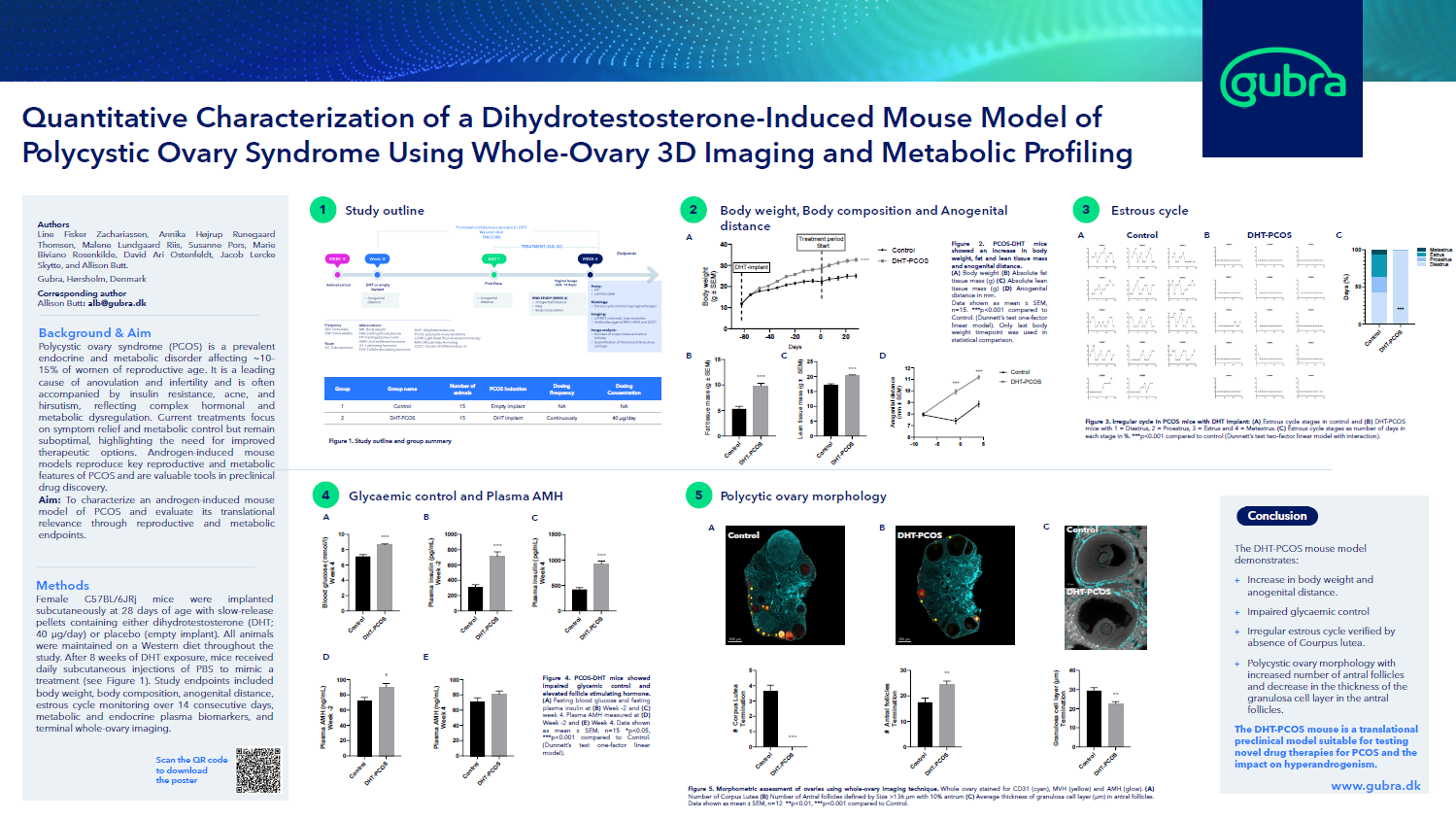
Gubra’s DHT-induced PMOS mouse model combines sustained exposure to the androgen dihydrotestosterone (DHT) with a Western diet to generate an integrated metabolic, endocrine, and ovarian phenotype. Metabolically, DHT-treated mice develop increased body weight and fat mass together with impaired glycemic control, including elevated fasting glucose and insulin.
Sustained DHT exposure provides the endocrine driver and induces persistent disruption of the estrous cycle. Whole-ovary 3D imaging further reveals the consequences for ovarian function, including an accumulation of antral follicles, an absence of corpora lutea, and reduced granulosa cell-layer thickness. Estrous cycle monitoring, circulating biomarkers, ovary histology, and follicle quantification can therefore be combined to assess responses across all three disease domains. This model is particularly relevant for anti-androgenic therapies, metabolic treatments, and candidates designed to address the three components of PMOS.
Letrozole-induced rat model: A leaner PMOS phenotype
The letrozole-induced rat model provides a complementary approach based on pharmacological aromatase inhibition, which reduces the conversion of androgens into estrogens. This endocrine imbalance suppresses estrous cycling during letrozole administration. Cycling resumes following withdrawal, demonstrating a hormonally driven and reversible reproductive phenotype. Ovarian function can be evaluated through estrous cycle monitoring, ovary histology, and whole-ovary 3D imaging of follicles, granulosa cells, and corpora lutea.
The model also supports metabolic profiling through measurements of body weight, body composition, fasting glucose, and plasma insulin. Under standard-diet conditions, letrozole increases body weight without disruption of fasting glycemic markers. This rat model therefore represents a comparatively lean PMOS phenotype and is particularly relevant for therapies targeting endocrine regulation, ovarian function, and follicular development. A sustained high-fat diet can be incorporated when a stronger metabolic phenotype is required.
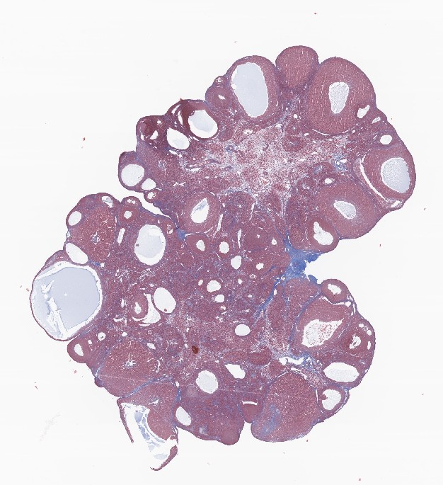
Ovary from rat stained with Masson’s Trichrome
Studying menopause and ovarian insufficiency in female rodent models
Loss of ovarian function affects far more than the reproductive system. Declining ovarian hormone production can alter body composition, metabolic regulation, skeletal integrity, and the structure of reproductive tissues. Complementary ovariectomy (OVX) and primary ovarian insufficiency (POI) models allow researchers to distinguish the systemic consequences of complete hormone loss from the effects of impaired ovarian function.
OVX rat model: Evaluating the systemic consequences of menopause
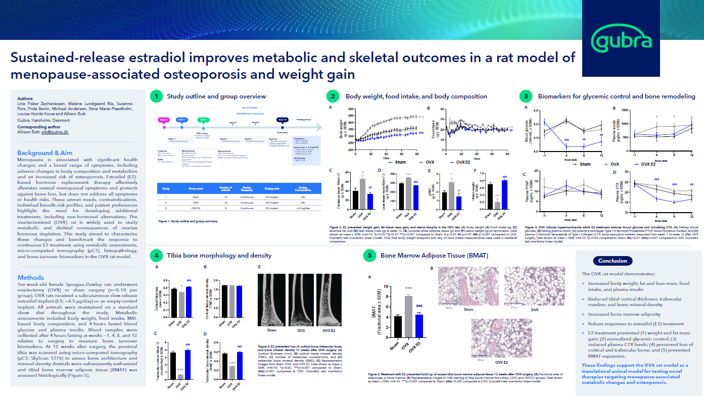
In the OVX rat model, surgical removal of the ovaries produces complete and controlled ovarian hormone deprivation. This robust and reproducible model is particularly useful for preclinical menopause research focused on the systemic consequences of estrogen loss, including menopause-associated weight gain, metabolic dysfunction, osteoporosis, and reproductive-tissue atrophy. Because the ovaries are absent, the OVX model is not suitable for assessing ovarian reserve or therapies intended to protect or restore ovarian function.
Relevant endpoints capture the effects of ovarian hormone loss across several interconnected systems such as:
- Metabolic endpoints: Ovarian hormone loss can increase food intake, body weight, adiposity, and insulin levels. These changes are reproduced in Gubra’s OVX rat model, providing endpoints for evaluating whether a therapy can limit postmenopausal weight gain, preserve healthy body composition, or improve metabolic regulation.
- Skeletal endpoints: Estrogen depletion accelerates bone turnover and progressively compromises bone structure. In the OVX rat, this includes reductions in trabecular bone number, bone mineral density, and cortical thickness, together with increased bone marrow adiposity. The ability of a therapy to prevent or reverse these changes can be evaluated using micro-CT, serum bone-turnover markers such as P1NP and CTX, and bone histology.
- Reproductive-tissue endpoints: Ovarian hormone loss causes uterine and vaginal atrophy, including reduced uterine weight, thinning of the vaginal epithelium, and changes in tissue structure. The ability of a therapy to prevent or reverse these effects provides clinically relevant endpoints for treatments targeting vulvovaginal atrophy and other genitourinary consequences of menopause. Additionally, circulating hormone levels, such as AMH or LH, are significantly altered due to the loss of ovaries.
By combining metabolic, skeletal, endocrine, and tissue-level readouts, the OVX rat model provides an integrated platform for evaluating therapies designed to reduce the systemic consequences of menopause.
Primary ovarian insufficiency mouse model and ovarian reserve assessment
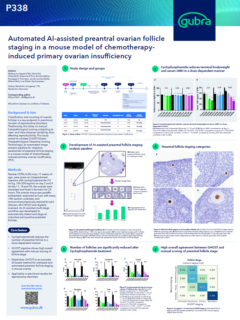
Gubra’s chemotherapy-induced POI model uses cyclophosphamide in mice to simulate the loss of ovarian reserve caused by cancer treatment, sometimes also called a model of chemically-induced menopause. Cyclophosphamide treatment reduces the ovarian follicle pool, with particularly pronounced depletion of primordial and primary follicles. Unlike the OVX model, the ovaries remain present, making this model suitable for evaluating compounds designed to protect the ovary, preserve or activate the remaining follicle pool, or promote follicle health following chemotherapy.
Ovarian function can be evaluated using complementary endocrine, histological, and whole-organ imaging endpoints:
- Ovarian reserve endpoints: Cyclophosphamide reduces circulating anti-Müllerian hormone (AMH) and depletes primordial and primary follicles. Serum AMH provides an endocrine measure of diminished ovarian reserve, while direct follicle quantification determines whether a therapy preserves the follicles that support future reproductive function.
- Follicle development and ovulatory endpoints: Two-dimensional ovary histology and whole-ovary 3D imaging can quantify and classify primordial, primary, secondary, antral, and preovulatory follicles. Quantification of corpora lutea provides an additional measure of ovulatory activity. Together, these endpoints show whether a compound protects the initial follicle pool and supports continued follicular development.
- Ovarian tissue-health endpoints: Chemotherapy can damage oocytes and surrounding ovarian tissue in addition to reducing follicle numbers. Markers of oocytes, granulosa cells, vasculature, and apoptosis can characterize the location and extent of this injury, while Picrosirius Red staining can assess ovarian fibrosis. These endpoints help determine whether a therapy protects the broader ovarian environment needed to support follicle survival and maturation.
By combining circulating AMH, ovary histology, follicle quantification, and whole-ovary 3D imaging, the POI mouse model provides an integrated assessment of ovarian protection and follicle health.
Studying cardiorenal disease in a female mouse model
The female ReninAAV UNx db/db mouse model
Cardiorenal disease is an important but sometimes overlooked component of women’s health. Approximately one in ten women worldwide lives with chronic kidney disease, which is closely connected to cardiovascular and metabolic risk. Biological sex can influence kidney disease development, progression, and treatment response, supporting the inclusion of female animal models in preclinical cardiorenal research.
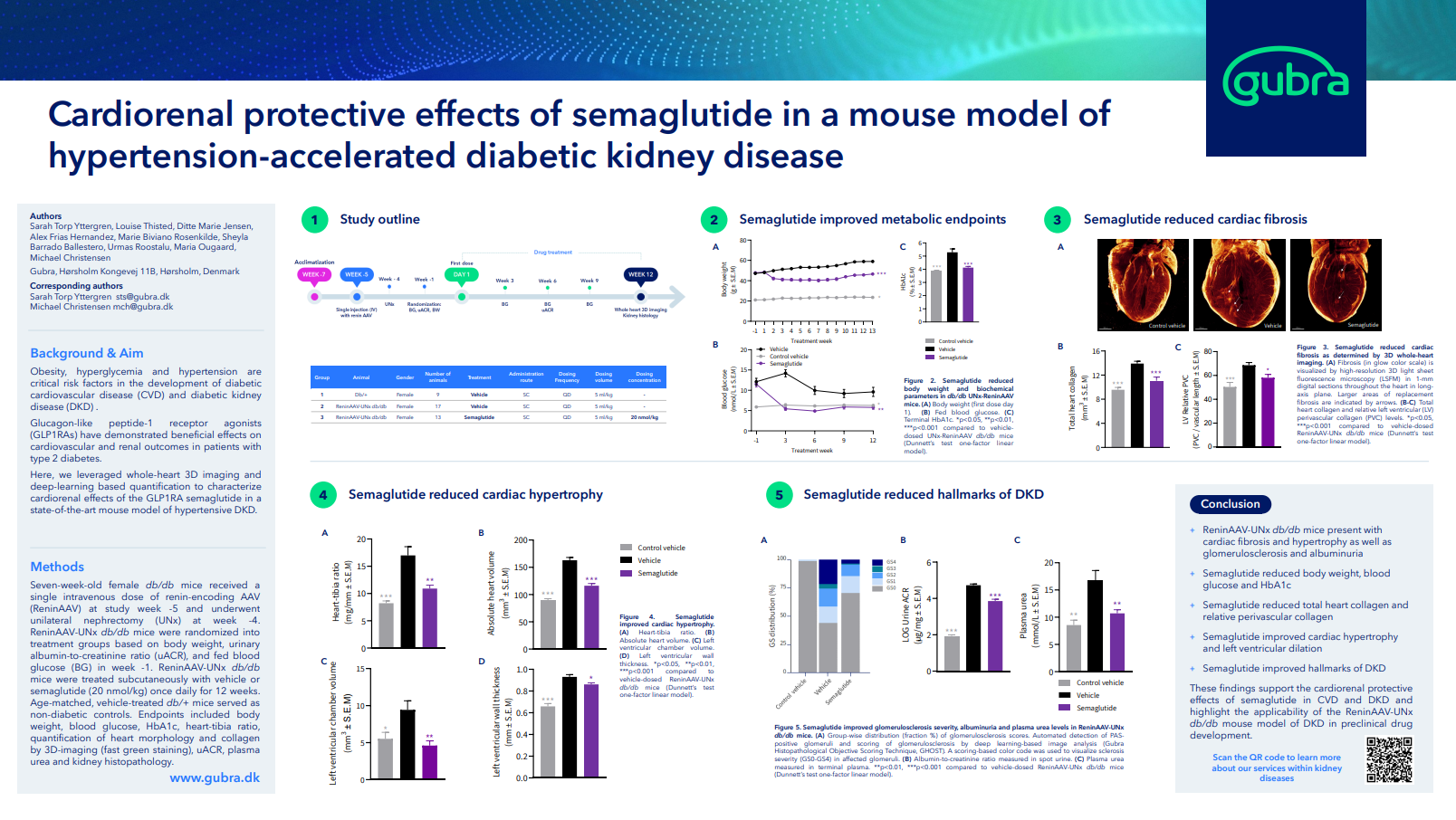
Gubra’s female diabetic kidney disease model (ReninAAV UNx db/db mouse model) combines obesity, hyperglycemia, hypertension, and renal injury to reproduce progressive diabetic kidney disease and its cardiovascular complications.
- Renal effects: The model develops progressive albuminuria, glomerulosclerosis, kidney fibrosis, tubular injury, and inflammation. Disease progression and therapeutic efficacy can be assessed using blood pressure, urine albumin-to-creatinine ratio, plasma urea, urinary KIM-1, AI-assisted histological scoring of glomulosclerosis, and markers of fibrosis, tubular injury, and inflammation. Bulk or single-nucleus RNA sequencing can provide additional insight into treatment effects on renal pathways and individual kidney cell types.
- Cardiac effects: Hypertension-accelerated diabetic kidney disease is accompanied by cardiac fibrosis and hypertrophic remodeling. Whole-heart 3D imaging enables spatial quantification of collagen burden across the whole heart and in 17 regions of the heart. Additionally, 3D imaging enables rapid measurements of heart-to-tibia ratio, heart volume, left ventricular chamber volume, and ventricular wall thickness to monitor compound effects on cardiac hypertrophy and structure.
Responsiveness to semaglutide alone and in combination with lisinopril demonstrates that the model can detect improvements across metabolic, cardiovascular, and renal endpoints. Learn more about the ReninAAV UNx db/db mouse model of diabetic kidney disease.
Technologies for evaluating female reproductive health in preclinical models
Female reproductive health cannot be characterized through a single measurement. Gubra combines longitudinal monitoring with metabolic, skeletal, histological, imaging, and molecular endpoints. Body weight, food intake, body composition, glucose, insulin, and circulating hormones can be monitored across models, while longitudinal micro-CT scanning assesses bone density and microarchitecture in the OVX rat model.
Estrous cycle and metabolic monitoring
Estrous cycles in animals are monitored through daily vaginal lavage followed by cytological evaluation of the collected cells. Changes in epithelial and immune-cell composition are used to classify samples as proestrus, estrus, metestrus, or diestrus. Gubra has automated this process using digital slide scanning and AI-assisted scoring, increasing throughput and reducing the variability associated with manual classification.
Longitudinal estrous cycle monitoring can identify persistent cycle arrest, irregular cycling, and the recovery of cyclicity following treatment or withdrawal of model-inducing agent. These data can be integrated with metabolic and endocrine measurements to determine how changes in reproductive function relate to the broader disease phenotype and treatment response.
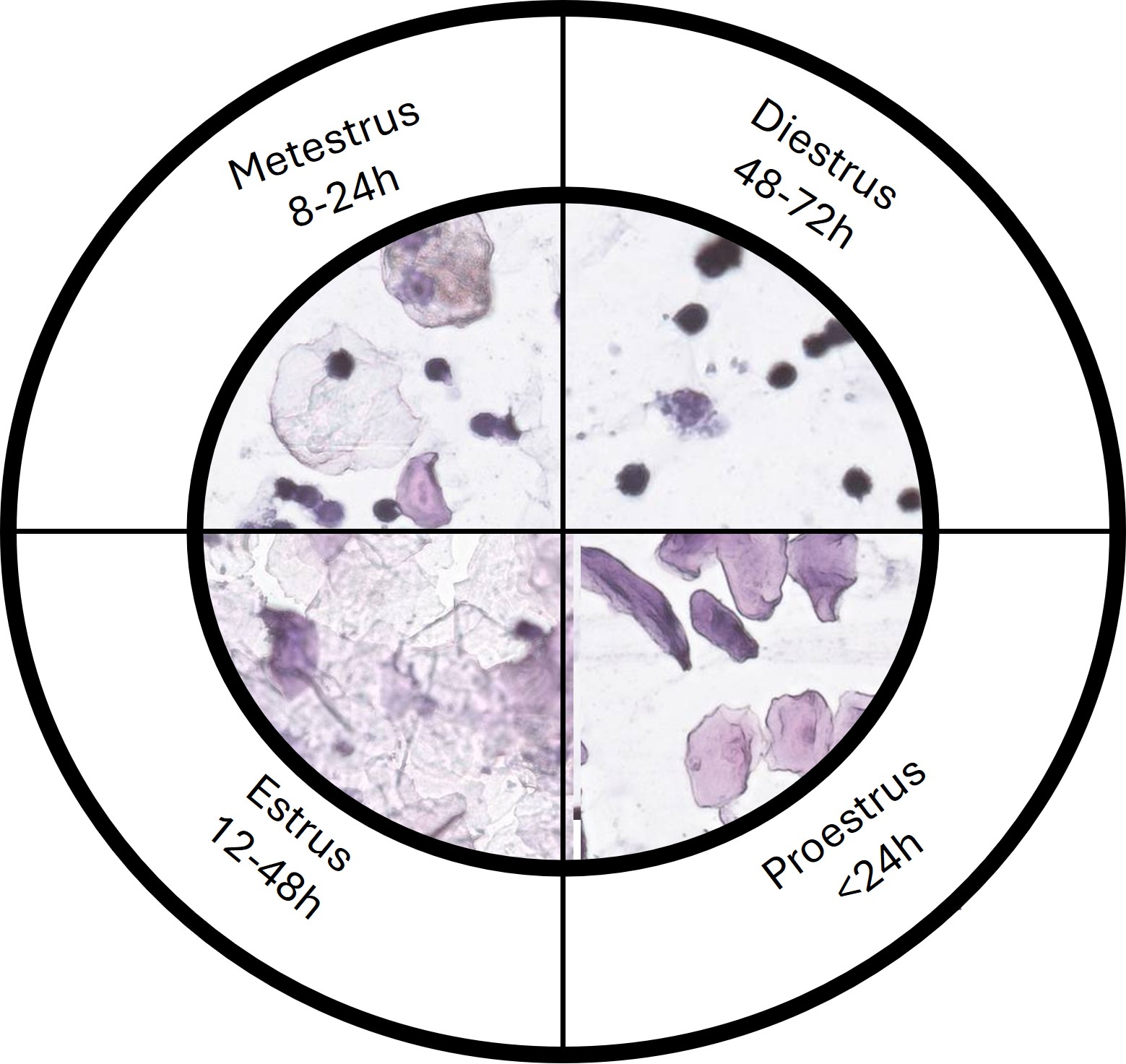
Example images of a full mouse estrous cycle
Whole-ovary 3D imaging and follicle quantification
Ovary histology and whole-ovary 3D imaging provide complementary methods for detecting, classifying, and quantifying follicles and oocytes. Measuring the number and distribution of follicles across developmental stages can reveal follicular arrest, impaired maturation, or depletion of the ovarian reserve.
- AI-assisted ovary histology: Traditional ovary histology provides cellular detail on follicle morphology, oocyte integrity, atresia, and surrounding tissue health. At Gubra, p63 staining is combined with AI-assisted image analysis to detect and stage preantral follicles across tissue sections. Automation increases throughput and reduces variability while retaining the tissue detail needed to evaluate treatment effects. Read more in our blog article Preclinical ovary histology: AI-assisted analysis of reproductive tissue and endpoints.
- Whole-ovary 3D imaging: Following tissue clearing and immunolabeling, light-sheet fluorescence microscopy can visualize and quantify follicles throughout the intact ovary. Follicles can be classified by developmental stage alongside measurements of corpora lutea, granulosa-layer thickness, vasculature, and cell death. Whole-organ analysis reduces sectioning bias and provides spatial information that cannot be obtained from selected two-dimensional sections.
Together, histology reveals the cellular condition of follicles and surrounding tissue, while whole-ovary 3D imaging shows how follicle number and distribution change across the entire organ.
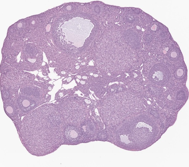
Ovary from mouse stained with Hematoxylin Eosin
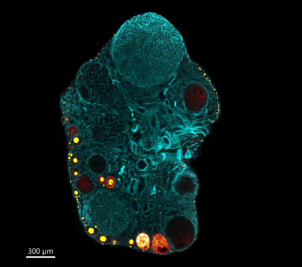
3D image of a mouse ovary
Discuss your preclinical women’s health study
Choosing a model and endpoints that match your research question is an important step in study design. Contact our sales team to discuss Gubra’s PCOS, POI, menopause, or female cardiorenal disease models here.
Frequently asked questions about preclinical women’s health research
Gubra, a preclinical women’s health CRO, offers two PMOS models: a DHT and Western diet-induced mouse model and a letrozole-induced rat model. Additional preclinical models include a cyclophosphamide-induced POI mouse model, an OVX menopause rat model, and a female ReninAAV UNx db/db cardiorenal mouse model.
Preclinical women’s health studies can include estrous cycle monitoring, circulating hormones, body composition, glucose regulation, ovarian reserve assessment, ovary histology, follicle and oocyte quantification, whole-ovary 3D imaging, bone microarchitecture, and RNA sequencing. Endpoints should be selected according to the disease model, therapeutic target, and go/no-go decisions the study must address.
Yes. Female models can evaluate metabolic, skeletal, renal, and cardiovascular diseases. Gubra’s female ReninAAV UNx db/db mouse model supports integrated assessment of diabetic kidney disease and cardiac remodeling, while the OVX rat captures metabolic and skeletal consequences of ovarian hormone loss.