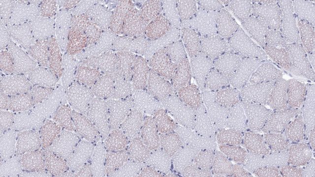
How can preclinical models support preclinical drug development for muscle diseases?
Muscle diseases and conditions encompass a diverse range of inherited, autoimmune, inflammatory, and degenerative disorders, as well as age-related sarcopenia and muscle wasting associated with chronic disease or disuse. These conditions can lead to loss of muscle mass and function, reduced mobility, and impaired quality of life, with some also compromising respiratory function. The underlying mechanisms are complex and, in many cases, incompletely understood, while effective treatment options remain limited. Drug development is further complicated by disease heterogeneity and the challenge of defining meaningful therapeutic endpoints. Translational preclinical models are therefore essential for elucidating disease mechanisms and evaluating novel therapies to preserve or restore muscle health. Advanced muscle histology can reveal structural, cellular, and pathological changes that are not captured by functional measures alone. These insights can guide candidate selection, dose optimization, and translational biomarker development throughout preclinical drug development.
What is skeletal muscle histology, and how can it support preclinical research?
Skeletal muscle histology reveals what muscle weight, body composition, and functional testing cannot: how muscle tissue is structured, maintained, and altered at the cellular level.
Skeletal muscle histology provides detailed insights into muscle health by revealing changes in muscle fiber size, number, and organization. Histological analysis can identify loss of muscle fiber integrity, shifts in fiber size distribution, and changes in the balance of slow- and fast-twitch fiber types. Histology also provides insight into structural and metabolic changes, including altered vascularization, lipid accumulation, glycogen storage, and glucose transport expression.
These readouts are particularly valuable in preclinical rodent models of sarcopenia, cachexia, and drug-induced muscle atrophy, where responses may differ considerably between individual muscles and fiber populations. Muscle histology can also support research into obesity, diabetes, and other metabolic diseases by determining whether weight-loss therapies preserve healthy muscle while reducing body weight.
When integrated with functional assessments and systemic metabolic endpoints, histology helps researchers determine whether a treatment preserves muscle architecture, changes muscle composition, or acts through a specific biological pathway in mouse and rat models. Importantly, these histological endpoints can also help elucidate the mode of action of novel drug candidates and differentiate their efficacy from that of current therapies.
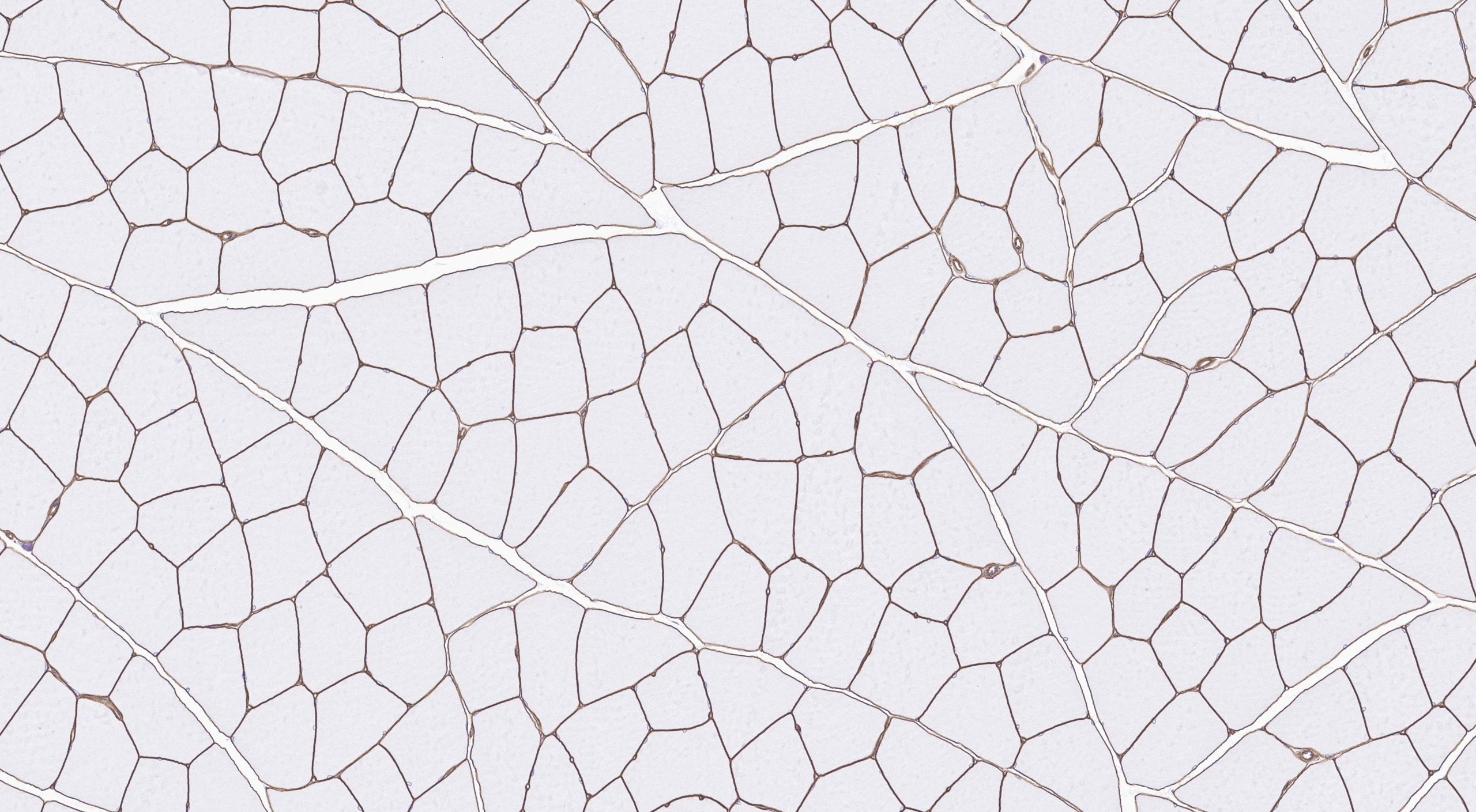
Cross-section of mouse gastrocnemius muscle stained for laminin. The brown laminin-positive staining outlines individual muscle fibers, enabling quantification of muscle fiber cross-sectional area (CSA), diameter, fiber number, and total tissue area.
Which skeletal muscle histology endpoints reveal muscle health and treatment response?
To evaluate complete muscle health in translational preclinical mouse models, combining structural and metabolic readouts can provide a holistic overview.
Muscle fiber size and type
The quantification and characterization of muscle fiber size and type can reveal whether muscle loss affects the entire fiber population or specific subsets. Laminin staining outlines individual fibers for quantification of muscle cross-sectional area (CSA), diameter, fiber number, and total tissue area.
Muscle fiber size provides a quantitative readout of skeletal muscle atrophy or hypertrophy in mice and rats. Muscle fiber typing adds key biological context, i.e. type I muscle fibers support sustained, oxidative activity, whereas type II fibers contract more rapidly, with type IIa combining fast contraction with substantial oxidative capacity and type IIb supporting predominantly glycolytic, powerful contractions.
Accordingly, slow-twitch muscles support endurance, while fast-twitch muscles provide quick, powerful movement. Immunofluorescent labelling of myosin heavy-chain isoforms (MYH7 for type I “slow-twitch” fibers, and MYH2 for type IIa and MYH4 for type IIb “fast-twitch” fibers) enables assessment of fiber-type distribution which, when combined with size measurements, reveals whether a model or treatment preferentially affects particular fiber populations. To address the technical demands of muscle fiber typing, Gubra has developed and optimized an in-house multiplex immunohistochemistry (IHC) platform, enabling simultaneous visualization of multiple fiber types within a single tissue section.
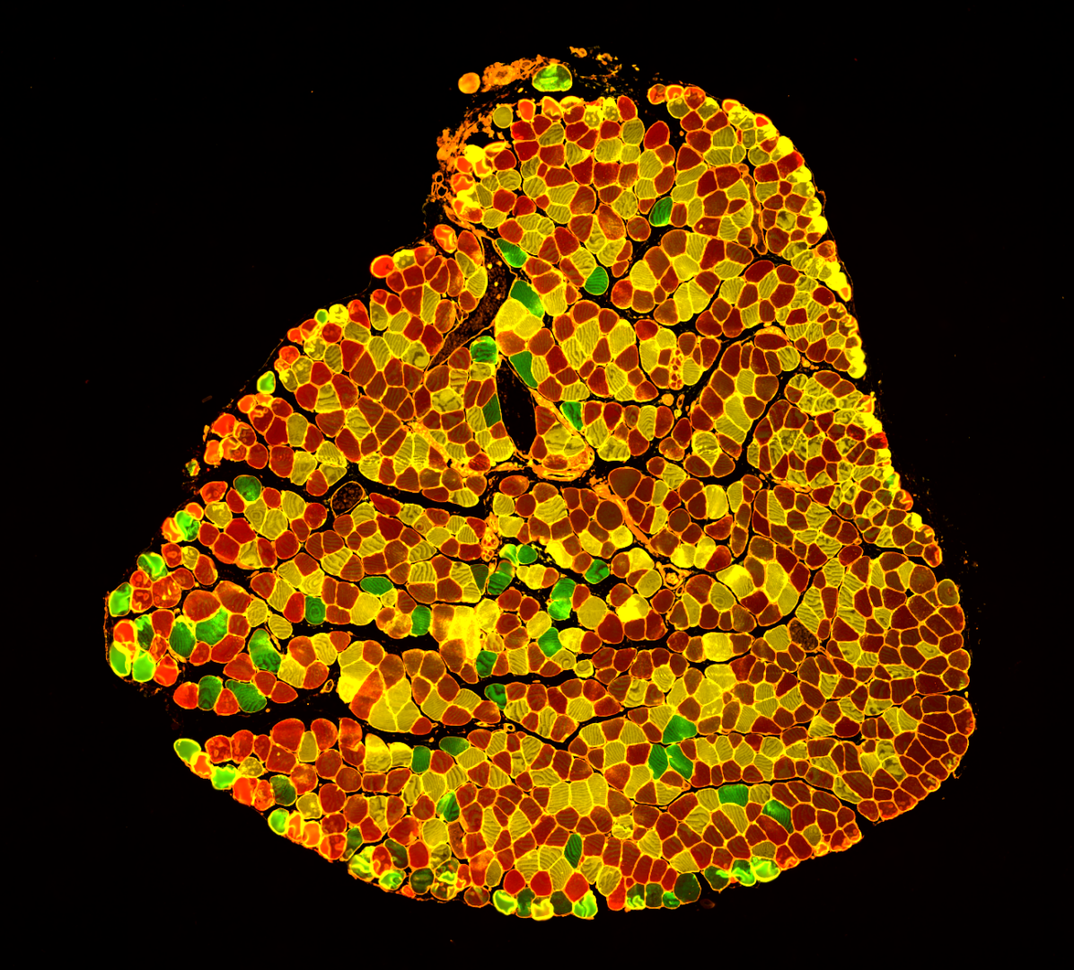
Cross-section of mouse soleus muscle stained to identify muscle fiber types. Each fiber type has a distinct color: yellow is fiber type I, red is muscle fiber type IIa, and green is muscle fiber type IIb.
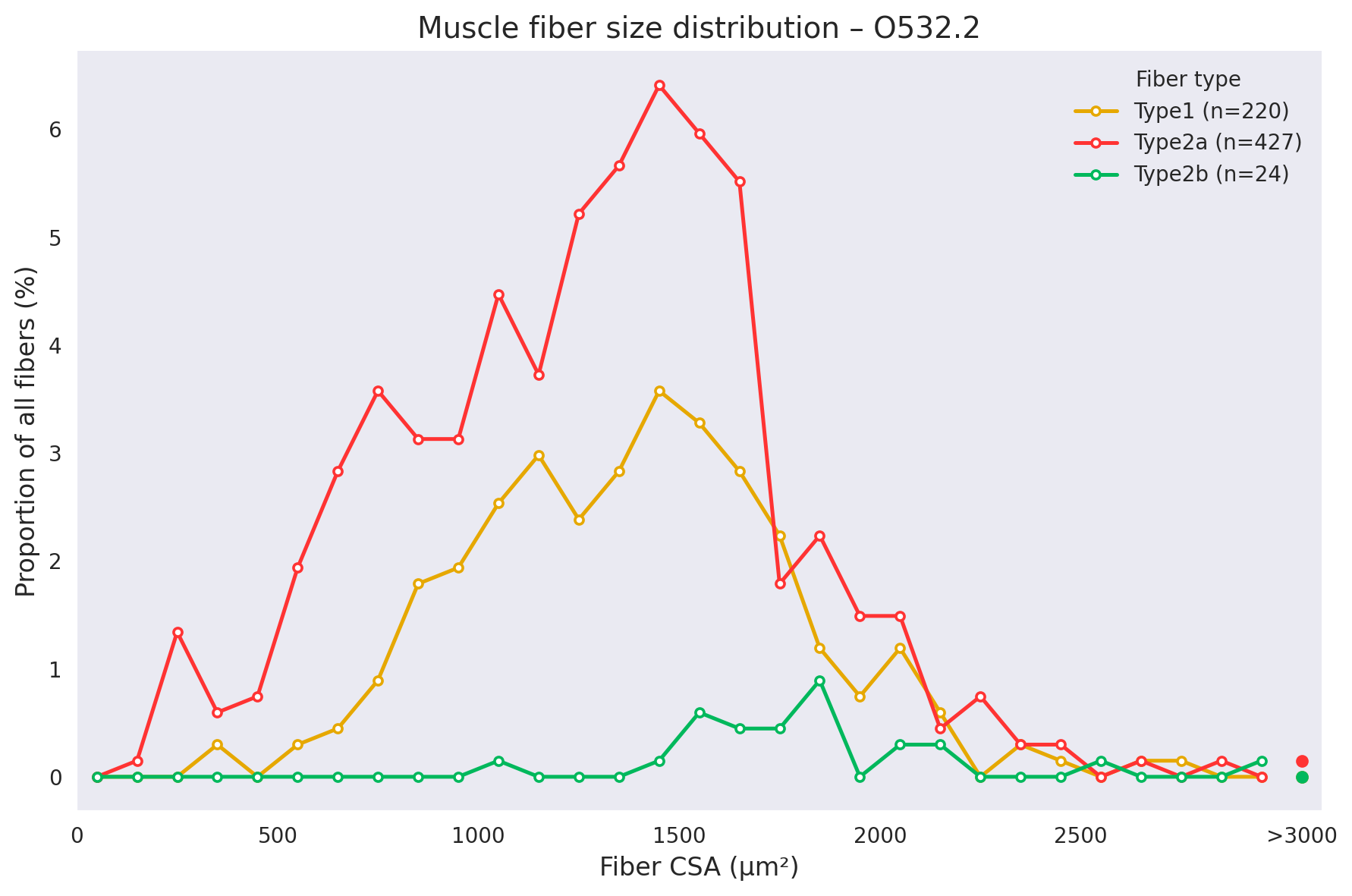
Subsequent digital image analysis enables quantitative assessment of muscle fiber type composition and fiber size distribution.
Fat accumulation within skeletal muscle
Skeletal muscle can accumulate intramuscular adipose tissue or lipid droplets within the fibers themselves, reflecting metabolic imbalances in obesity-associated diseases. Lipid infiltration in skeletal muscle is commonly associated with reduced insulin sensitivity, inflammation, and reduced muscle quality. These effects are recapitulated in rodent models of diet-induced obesity and muscle wasting.
Hematoxylin and eosin (H&E) staining reveals and allows for quantification of intramuscular adipose tissue, while Oil Red O (ORO) staining detects intramyocellular lipids. Quantifying the extent of adipose/lipid accumulation inside and between muscle fibers helps determine whether an intervention improves muscle quality in addition to preserving muscle size in preclinical animal models.
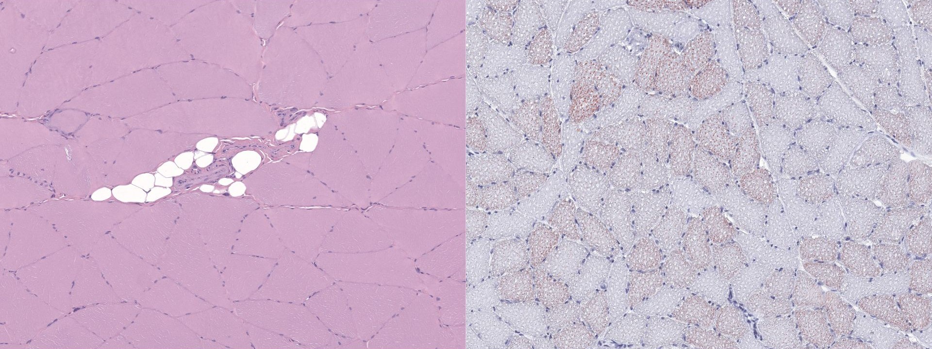
(Left) Cross-section of mouse quadriceps muscle. Stained with H&E. In the center an area with white intramuscular adipose tissue is visible. (Right) Cross-section of mouse quadriceps muscle. Stained with ORO. Dispersed red colored cells are muscle fibers containing intramyocellular lipids.
Glucose uptake and glycogen storage
Skeletal muscle is a major site of glucose metabolism, and glucose storage, making muscle histology particularly relevant in preclinical mouse models of obesity and diabetes. Periodic Acid-Schiff (PAS) staining visualizes glycogen deposition, while glucose transporter type 4 (GLUT4) staining provides insight into the molecular machinery responsible for insulin-stimulated glucose transport. When combined with the measurements described above, such as muscle fiber size and type, these muscle glucose metabolic endpoints provide a more complete view of how a drug candidate simultaneously affects muscle structure and metabolic function in rodent models of obesity and obesity-related complications.
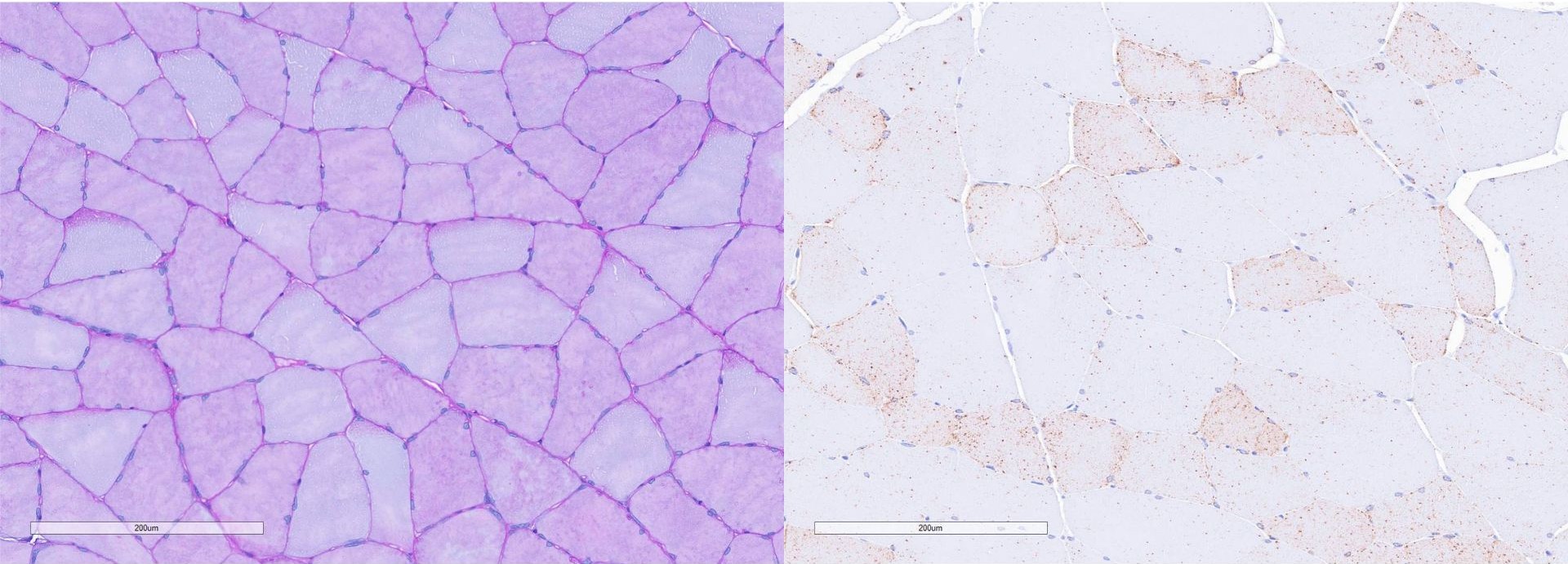
(Left) Cross-section of rat gastrocnemius muscle stained with PAS. Dispersed red colored cells contain glycogen. (Right) Cross-section of rat gastrocnemius muscle stained with GLUT4.
How is advanced histology applied in models of muscle atrophy, sarcopenia, and cachexia?
Advanced histology helps characterize how sarcopenic or cachexic mouse models have altered tissue structure, composition, and metabolism. In preclinical studies, quantitative histological endpoints can reveal whether a reduction in muscle mass reflects smaller fibers, loss of fibers, or a shift in fiber-type composition. These tissue-level changes provide biological context for concurrent changes in body composition, terminal muscle weight, and muscle strength endpoints.
In models of muscle atrophy, such as dexamethasone-induced muscle atrophy, laminin staining is useful for measuring reductions in fiber CSA and diameter. Fiber typing can then determine whether the atrophic response preferentially affects specific fiber populations. In sarcopenia research, broader panels can assess features associated with declining muscle quality, including fiber atrophy, altered fiber-type distribution, reduced vascularization, mitochondrial changes, and increased fat accumulation.
Cachexia involves loss of both body weight and muscle tissue, often alongside changes in energy balance and metabolism. In mouse models, such as cisplatin-induced cachexia, histology can determine how this systemic response affects individual muscle fibers and tissue composition. Assessment of intramuscular fat, glucose transport, and glycogen storage can further identify metabolic alterations that may contribute to impaired muscle function.
Combining quantitative histology with functional testing and intact whole-hindlimb 3D imaging provides a more complete assessment of treatment response. Whole-limb analysis shows where muscle loss occurs, while histology explains the cellular and metabolic changes underlying that loss. Functional testing quantifies the muscle capacity and function ex vivo. Together, these approaches can determine whether a candidate preserves muscle volume, maintains fiber quality, and supports functional recovery.
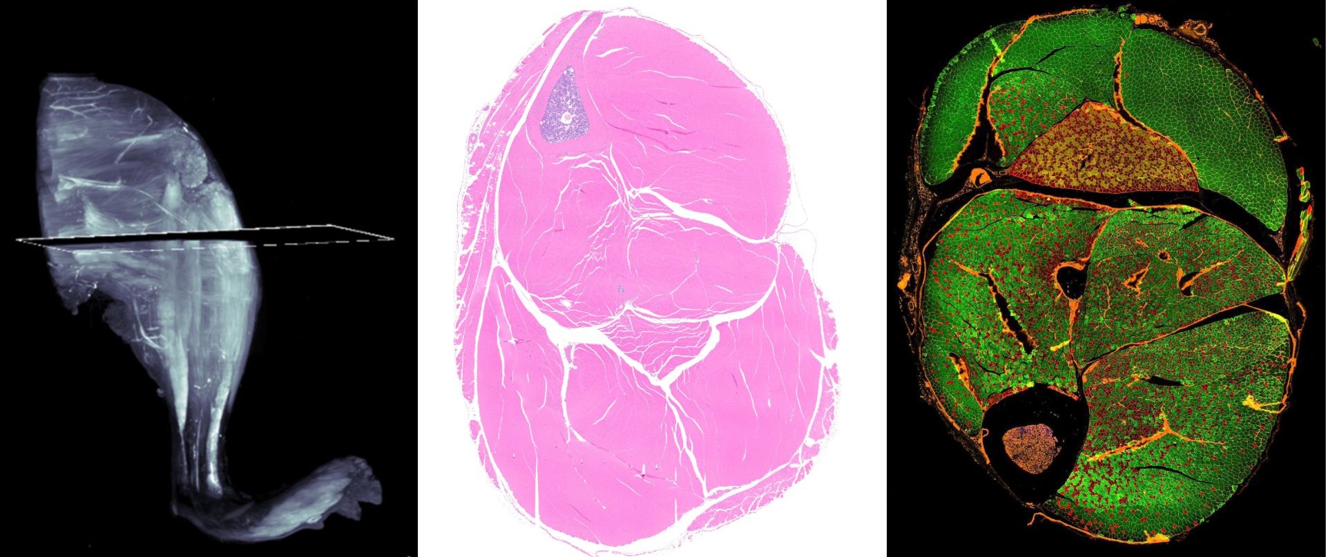
3D whole leg and 2D section of the whole leg representing several muscle types. (Left) 3D whole leg maximum projection from light sheet microscopy, (Center) cross section of whole leg stained with H&E, and (Right) muscle fiber type immunofluorescent staining.
Why combine 2D muscle histology with 3D imaging and functional endpoints?
At Gubra, we combine whole-hindlimb 3D imaging, 2D histology, in vivo grip strength, and ex vivo contractility measurements to evaluate muscle health and function in our preclinical models. Together, these complementary approaches connect changes in muscle structure with functional outcomes to characterize treatment response.
- Quantitative 3D imaging measures muscle volume and morphology across the intact hindlimb, reducing the sampling limitations associated with muscle dissection and weighing. Keeping the leg intact also preserves the anatomical relationships among individual muscles, bones, and other structures, allowing treatment effects to be evaluated within their broader anatomical context.
- Quantitative 2D histology adds cellular detail to help interpret changes detected by 3D imaging. Measurements of fiber size and fiber-type composition help characterize muscle remodeling, while dedicated stains can reveal fat accumulation and metabolic features that volumetric measurements alone cannot resolve.
- Functional endpoints link muscle structure to performance. In vivo grip strength testing assesses the force generated during a gripping task, providing an integrated measure of neuromuscular performance. Complementary ex vivo assays can measure contractile force, contraction dynamics, fatigue, and recovery in isolated muscles. Subsequent histological analysis of the tested muscles can help determine whether functional differences are associated with changes in fiber size or fiber-type composition.
Integrating these diverse readouts connects muscle quantity with muscle quality and function. Whole-limb imaging identifies where treatment effects occur, our advanced histology platform provides data to interpret the underlying tissue structural changes, and functional testing establishes whether those changes translate into improved muscle performance. This combined approach provides a complete assessment of therapeutic efficacy in mouse and rat models of muscle atrophy, sarcopenia, cachexia, and more.
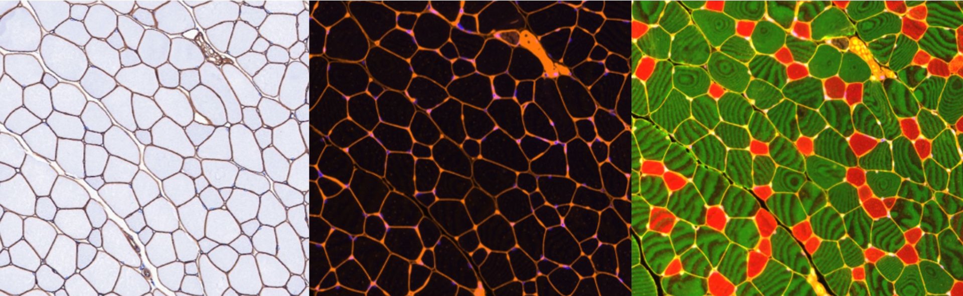
Section of muscle stained for (Left) laminin, (Center) Wheat germ agglutinin (WGA), and (Right) muscle fiber type. By using the laminin and WGA stains, we can delineate individual muscle fibers, allowing for quantification of changes in different muscle fiber types.
We are further expanding our muscle histology portfolio into mitochondrial assessment, including COX4- and TOMM20-based staining approaches, to provide insights into mitochondrial content, organization, and bioenergetic adaptations in skeletal muscle.
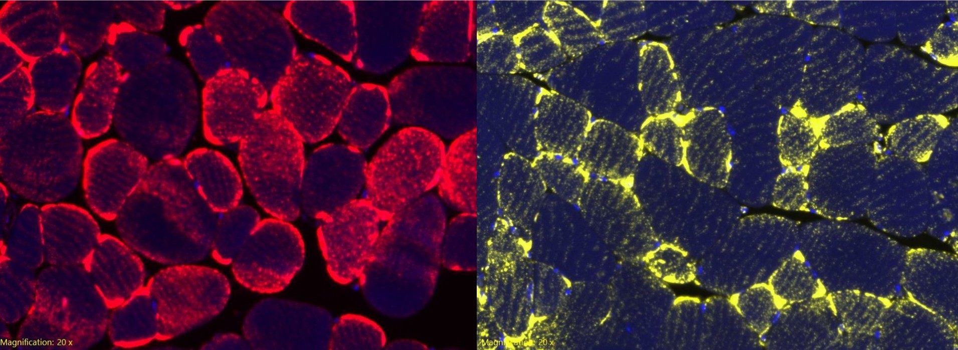
Section of mouse muscle stained with COX4 in red (Left) and TOMM20 in yellow (Right).
In parallel, we are developing automated image analysis workflows to quantify myonuclear relocation patterns, a key histological indicator of muscle regeneration and repair.
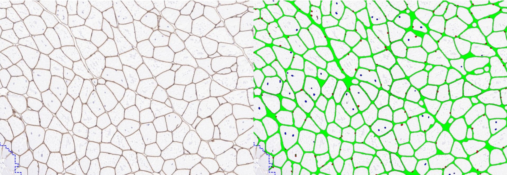
(Left) Brown laminin staining is used to delineate muscle fibers, and blue hematoxylin staining is used to mark the cell nuclei. (Right) Using image analysis, we can distinguish between peripheral nuclei (red) located under the cell membrane (green) and centralized nuclei (blue) located in the middle of the muscle fiber.
Preclinical muscle histology at Gubra
At Gubra, preclinical muscle histology can be used as a stand-alone analysis or combined with whole-hindlimb 3D imaging, in vivo assessments and ex vivo muscle function testing. Across mouse and rat models of muscle atrophy, sarcopenia, cachexia and metabolic disease, these complementary endpoints provide quantitative insights into muscle structure, composition, metabolism and treatment response. Explore our preclinical muscle research platform or contact our team to discuss the most relevant endpoints for your next preclinical study.
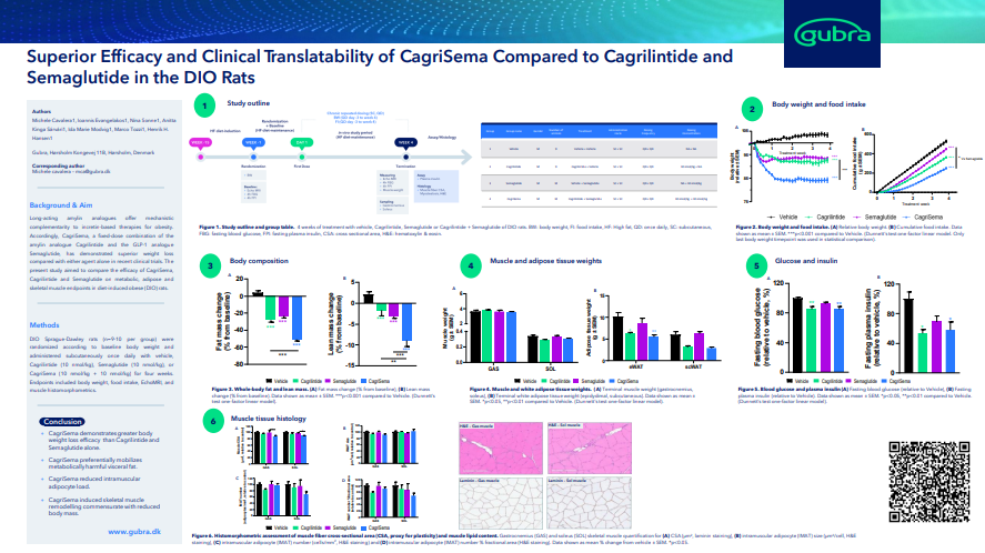
Superior Efficacy and Clinical Translatability of CagriSema Compared to Cagrilintide and Semaglutide in the DIO rats
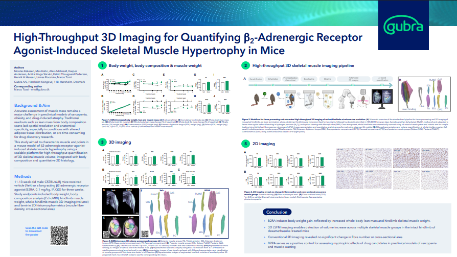
High-Throughput 3D Imaging for Quantifying b2-Adrenergic Receptor Agonist-Induced Skeletal Muscle Hypertrophy in Mice
Frequently asked questions about skeletal muscle histology
What can skeletal muscle histology measure?
Muscle fiber size, muscle fiber type, myosteatosis, glycogen deposition, glucose transport, mitochondrial biology.
How is muscle fiber cross-sectional area measured?
The muscle samples are stained with laminin or WGA to determine the size of muscle fibers.
Which stains and markers are used in skeletal muscle histology?
Laminin is used to evaluate muscle fiber size and muscle mass. CD31 represents vascularization. H&E and ORO stain are used to evaluate myosteatosis. PAS staining is used to quantify glycogen deposition. Anti-GLUT4 IHC is performed to evaluate glucose transport. MYH7 (Type I), MYH2 (Type IIa), and MYH4 (Type IIb) are used for muscle fiber typing. Finally, COX4- and TOMM20-based stains provide insights into mitochondrial biology.
Can we distinguish type I and type II muscle fibers?
Yes, we use different immunofluorescent antibodies specific to the two muscle fiber types and quantify the number of each type.